Clinical value and applicability of radiomics in differential diagnosis of dual-phenotype hepatocellular carcinoma and intrahepatic cholangiocarcinoma.
IF 1.5 Q3 RADIOLOGY, NUCLEAR MEDICINE & MEDICAL IMAGING
Chen-Cai Zhang, Da Lu, Jun Yang, Ling Zhang, Xia-Feng Zeng, Xiang-Ming Fang, Cun-Geng Fan
{"title":"Clinical value and applicability of radiomics in differential diagnosis of dual-phenotype hepatocellular carcinoma and intrahepatic cholangiocarcinoma.","authors":"Chen-Cai Zhang, Da Lu, Jun Yang, Ling Zhang, Xia-Feng Zeng, Xiang-Ming Fang, Cun-Geng Fan","doi":"10.4329/wjr.v17.i6.108247","DOIUrl":null,"url":null,"abstract":"<p><strong>Background: </strong>Dual-phenotype hepatocellular carcinoma (HCC) is a relatively new subtype of HCC. Studies have shown that in the context of chronic hepatitis, liver cirrhosis, and other liver conditions, some intrahepatic cholangiocarcinomas (ICCs) exhibit an enhancement pattern similar to that of HCC. Both dual-phenotype HCC (DPHCC) and ICC can express biliary markers, making imaging and pathology differentiation difficult. Currently, radiomics is widely used in the differentiation, clinical staging, and prognosis assessment of various diseases. Radiomics can effectively differentiate DPHCC and ICC preoperatively.</p><p><strong>Aim: </strong>To evaluate the value of radiomics in the differential diagnosis of DPHCC and ICC and to validate its clinical applicability.</p><p><strong>Methods: </strong>In this retrospective study, the data of 53 DPHCC patients and 124 ICC patients were collected retrospectively and randomly divided into training and testing sets at a ratio of 7: 3. After delineation of regions of interest and feature extraction and selection, radiomics models were constructed. Receiver operating characteristic curve analysis was conducted to calculate the area under the curve (AUC) for each model. The AUC values of radiologists with and without assistance from the model were also assessed.</p><p><strong>Results: </strong>In the training set, the AUC value of the radiomic model was the highest, and the combined model and the radiomic model had similar AUC (<i>P</i> > 0.05); the differences in the AUC values between the combined model and the clinical-sign model was statistically significant (<i>P</i> < 0.05). In the testing set, the AUC value of the combined model was the highest, and the differences in the AUC values between the combined model and the clinical-sign model was statistically significant (<i>P</i> < 0.05). With model assistance, the AUC values of Doctor D (10 years of experience in abdominal imaging diagnosis) and Doctor E (5 years of experience in abdominal imaging diagnosis) both increased.</p><p><strong>Conclusion: </strong>Radiomics can differentiate DPHCC and ICC, and with assistance from the developed model, the accuracy of less experienced doctors in the differential diagnosis of these two diseases can be improved.</p>","PeriodicalId":23819,"journal":{"name":"World journal of radiology","volume":"17 6","pages":"108247"},"PeriodicalIF":1.5000,"publicationDate":"2025-06-28","publicationTypes":"Journal Article","fieldsOfStudy":null,"isOpenAccess":false,"openAccessPdf":"https://www.ncbi.nlm.nih.gov/pmc/articles/PMC12210198/pdf/","citationCount":"0","resultStr":null,"platform":"Semanticscholar","paperid":null,"PeriodicalName":"World journal of radiology","FirstCategoryId":"1085","ListUrlMain":"https://doi.org/10.4329/wjr.v17.i6.108247","RegionNum":0,"RegionCategory":null,"ArticlePicture":[],"TitleCN":null,"AbstractTextCN":null,"PMCID":null,"EPubDate":"","PubModel":"","JCR":"Q3","JCRName":"RADIOLOGY, NUCLEAR MEDICINE & MEDICAL IMAGING","Score":null,"Total":0}
引用次数: 0
Abstract
Background: Dual-phenotype hepatocellular carcinoma (HCC) is a relatively new subtype of HCC. Studies have shown that in the context of chronic hepatitis, liver cirrhosis, and other liver conditions, some intrahepatic cholangiocarcinomas (ICCs) exhibit an enhancement pattern similar to that of HCC. Both dual-phenotype HCC (DPHCC) and ICC can express biliary markers, making imaging and pathology differentiation difficult. Currently, radiomics is widely used in the differentiation, clinical staging, and prognosis assessment of various diseases. Radiomics can effectively differentiate DPHCC and ICC preoperatively.
Aim: To evaluate the value of radiomics in the differential diagnosis of DPHCC and ICC and to validate its clinical applicability.
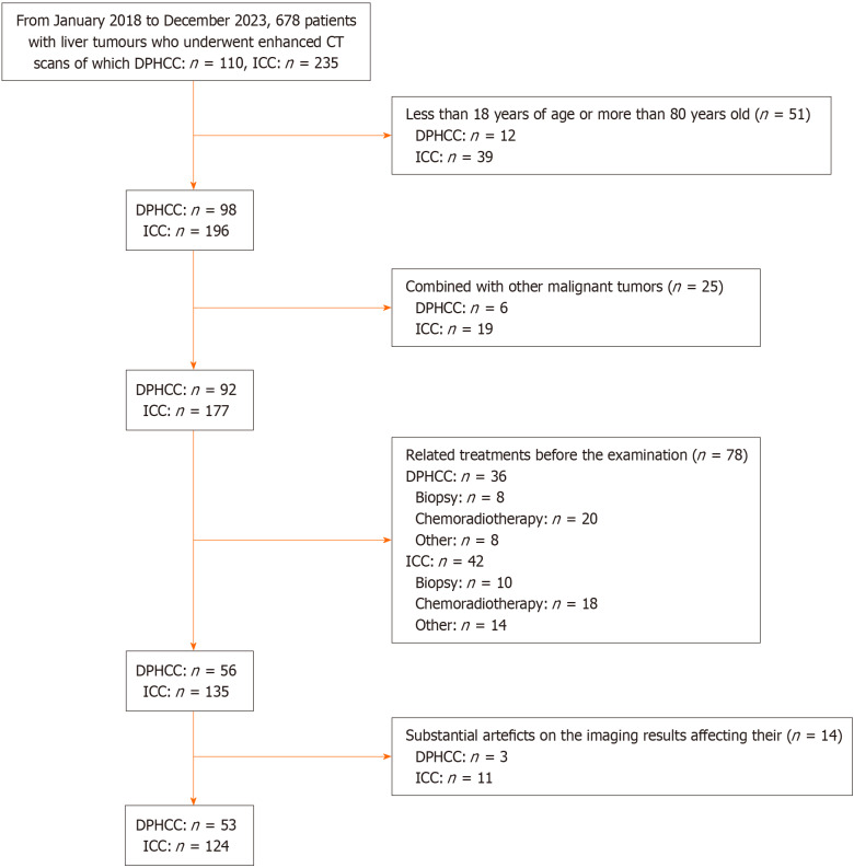
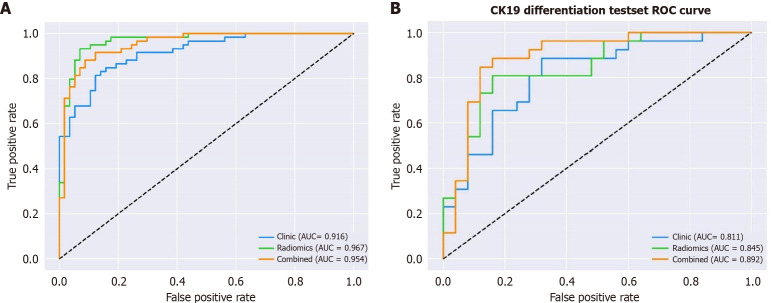
Methods: In this retrospective study, the data of 53 DPHCC patients and 124 ICC patients were collected retrospectively and randomly divided into training and testing sets at a ratio of 7: 3. After delineation of regions of interest and feature extraction and selection, radiomics models were constructed. Receiver operating characteristic curve analysis was conducted to calculate the area under the curve (AUC) for each model. The AUC values of radiologists with and without assistance from the model were also assessed.
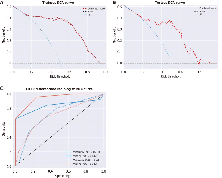
Results: In the training set, the AUC value of the radiomic model was the highest, and the combined model and the radiomic model had similar AUC (P > 0.05); the differences in the AUC values between the combined model and the clinical-sign model was statistically significant (P < 0.05). In the testing set, the AUC value of the combined model was the highest, and the differences in the AUC values between the combined model and the clinical-sign model was statistically significant (P < 0.05). With model assistance, the AUC values of Doctor D (10 years of experience in abdominal imaging diagnosis) and Doctor E (5 years of experience in abdominal imaging diagnosis) both increased.
Conclusion: Radiomics can differentiate DPHCC and ICC, and with assistance from the developed model, the accuracy of less experienced doctors in the differential diagnosis of these two diseases can be improved.

 求助内容:
求助内容: 应助结果提醒方式:


