Edward Godbold, Connor Luck, Camille Johnson, Ashley Disantis, Craig Mauro, Michael McClincy, William Anderst
{"title":"Using Dynamic Joint Space During Physiological Loading to Objectively Measure Hip Stability.","authors":"Edward Godbold, Connor Luck, Camille Johnson, Ashley Disantis, Craig Mauro, Michael McClincy, William Anderst","doi":"10.1002/jor.70011","DOIUrl":null,"url":null,"abstract":"<p><p>An objective measurement of hip stability during functional loading is needed to improve diagnosis, guide treatment decisions, and evaluate intervention success. This study aimed to develop a reference measurement for stable hips based upon dynamic minimum hip joint space (HJS) in healthy young adults. Synchronized biplane radiographs of the hips of 24 healthy young adults were collected (50 images/s) during treadmill walking and squatting. Subject-specific femur and pelvis models were created from CT scans, and bone motion was determined by a validated volumetric model-based tracking technique. The distance between femur and acetabulum subchondral bone surfaces was calculated during each movement. Range in minimum subchondral bone distance was measured in radial and sagittal regions of the acetabula. Regression analysis identified kinematics and morphologic predictors of range in minimum HJS. Range in minimum HJS during gait in the anterior-inferior (1.8 mm) and posterior-superior regions (1.7 mm) was 31%-38% larger than in the anterior-superior and superior regions (1.3 mm; p ≤ 0.001), and 13%-20% larger than in the posterior-inferior region (1.5 mm; p ≤ 0.001). No differences in minimum HJS were identified in radial regions during squat (range: 0.7-0.9 mm). No sex differences were identified. Femur head translation during gait was a stronger predictor of the range in minimum HJS than changes in femur head morphology. This suggests anterior-inferior to posterior-superior pistoning of the femur may be a mechanical mechanism for commonly observed pathology. These results suggest that gait is a better activity than squatting to assess dynamic hip stability when using this metric.</p>","PeriodicalId":16650,"journal":{"name":"Journal of Orthopaedic Research®","volume":" ","pages":"1666-1672"},"PeriodicalIF":2.3000,"publicationDate":"2025-09-01","publicationTypes":"Journal Article","fieldsOfStudy":null,"isOpenAccess":false,"openAccessPdf":"https://www.ncbi.nlm.nih.gov/pmc/articles/PMC12329620/pdf/","citationCount":"0","resultStr":null,"platform":"Semanticscholar","paperid":null,"PeriodicalName":"Journal of Orthopaedic Research®","FirstCategoryId":"3","ListUrlMain":"https://doi.org/10.1002/jor.70011","RegionNum":3,"RegionCategory":"医学","ArticlePicture":[],"TitleCN":null,"AbstractTextCN":null,"PMCID":null,"EPubDate":"2025/7/2 0:00:00","PubModel":"Epub","JCR":"Q2","JCRName":"ORTHOPEDICS","Score":null,"Total":0}
引用次数: 0
Abstract
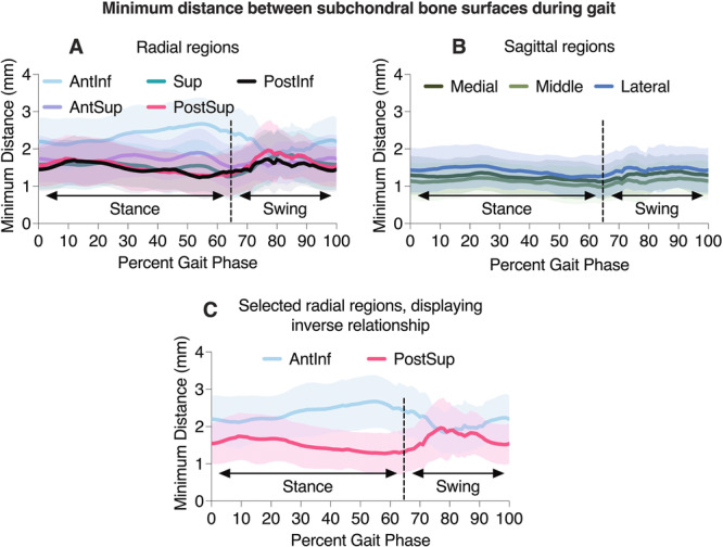
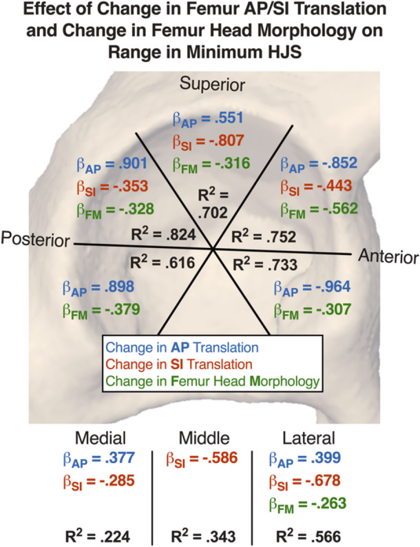
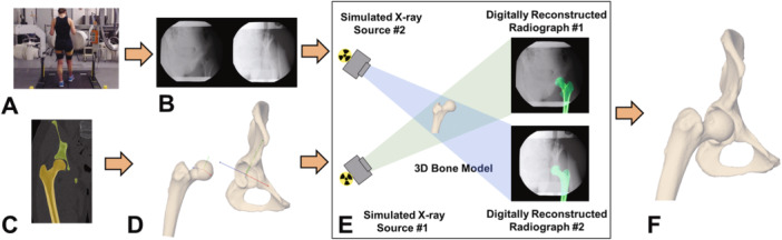
An objective measurement of hip stability during functional loading is needed to improve diagnosis, guide treatment decisions, and evaluate intervention success. This study aimed to develop a reference measurement for stable hips based upon dynamic minimum hip joint space (HJS) in healthy young adults. Synchronized biplane radiographs of the hips of 24 healthy young adults were collected (50 images/s) during treadmill walking and squatting. Subject-specific femur and pelvis models were created from CT scans, and bone motion was determined by a validated volumetric model-based tracking technique. The distance between femur and acetabulum subchondral bone surfaces was calculated during each movement. Range in minimum subchondral bone distance was measured in radial and sagittal regions of the acetabula. Regression analysis identified kinematics and morphologic predictors of range in minimum HJS. Range in minimum HJS during gait in the anterior-inferior (1.8 mm) and posterior-superior regions (1.7 mm) was 31%-38% larger than in the anterior-superior and superior regions (1.3 mm; p ≤ 0.001), and 13%-20% larger than in the posterior-inferior region (1.5 mm; p ≤ 0.001). No differences in minimum HJS were identified in radial regions during squat (range: 0.7-0.9 mm). No sex differences were identified. Femur head translation during gait was a stronger predictor of the range in minimum HJS than changes in femur head morphology. This suggests anterior-inferior to posterior-superior pistoning of the femur may be a mechanical mechanism for commonly observed pathology. These results suggest that gait is a better activity than squatting to assess dynamic hip stability when using this metric.
期刊介绍:
The Journal of Orthopaedic Research is the forum for the rapid publication of high quality reports of new information on the full spectrum of orthopaedic research, including life sciences, engineering, translational, and clinical studies.


 求助内容:
求助内容: 应助结果提醒方式:


