Jasmin Ordobazari, Charlotte Pfeiffer, Adriano Wang Leandro, Ina Quadflieg, Holger A Volk, Georga T Karbe
{"title":"Percutaneous cholecystostomy drain placement in cats: feasibility and safety of an ultrasound- and fluoroscopy-guided method.","authors":"Jasmin Ordobazari, Charlotte Pfeiffer, Adriano Wang Leandro, Ina Quadflieg, Holger A Volk, Georga T Karbe","doi":"10.1177/1098612X251336702","DOIUrl":null,"url":null,"abstract":"<p><p>ObjectivesThe aim of the present study was to evaluate the feasibility and safety of percutaneous ultrasound- and fluoroscopy-guided cholecystostomy drain placement.MethodsAn experimental cadaveric study was conducted on 16 cat cadavers weighing between 2.5 and 6.4 kg. Two drain systems were tested for percutaneous ultrasound- and fluoroscopy-guided placement: the nephrostomy component of a subcutaneous urethral bypass system (SUB-nephrostomy drain) and a paediatric percutaneous access set (paediatric-nephrostomy drain). Ultrasound-guided cholecystocentesis was performed via the 8th-12th intercostal space. Using a Seldinger technique, a guidewire was advanced into the gallbladder over which the drains were then passed under fluoroscopic control. Protocol modification was required mid experiment. Gallbladders were filled via catheterisation of the common bile duct before cholecystocentesis. After fluoroscopy-confirmed cholecystostomy drain placement, CT scans were performed to assess drain position, iatrogenic organ injuries and leakage. Leak pressure testing was performed followed by anatomic dissection. Organ injuries were recorded and classified as minor, moderate or severe.ResultsSUB-nephrostomy drain placement was performed in 15 cats and placement into the gallbladder was feasible in two: one was passed before and one after technique modification. Paediatric-nephrostomy drain placement was tested in one cat. The gallbladder could not accommodate the drain size, placement was not feasible and the device was not further tested. A CT scan of the two cats with drain placement showed a moderate amount of free peritoneal contrast, no pleural space penetration and one liver injury. Leakage occurred at a pressure of 4.5 cm H<sub>2</sub>O. For all drains, injuries recorded during anatomic dissection were to the liver, pleural space and gallbladder. The majority of injuries were classified as minor.Conclusions and relevancePercutaneous placement of cholecystostomy drains was not feasible with the method and devices tested. Further studies are needed to investigate alternative techniques in cats.</p>","PeriodicalId":15851,"journal":{"name":"Journal of Feline Medicine and Surgery","volume":"27 7","pages":"1098612X251336702"},"PeriodicalIF":2.1000,"publicationDate":"2025-07-01","publicationTypes":"Journal Article","fieldsOfStudy":null,"isOpenAccess":false,"openAccessPdf":"https://www.ncbi.nlm.nih.gov/pmc/articles/PMC12227865/pdf/","citationCount":"0","resultStr":null,"platform":"Semanticscholar","paperid":null,"PeriodicalName":"Journal of Feline Medicine and Surgery","FirstCategoryId":"97","ListUrlMain":"https://doi.org/10.1177/1098612X251336702","RegionNum":2,"RegionCategory":"农林科学","ArticlePicture":[],"TitleCN":null,"AbstractTextCN":null,"PMCID":null,"EPubDate":"2025/7/3 0:00:00","PubModel":"Epub","JCR":"Q2","JCRName":"VETERINARY SCIENCES","Score":null,"Total":0}
引用次数: 0
Abstract
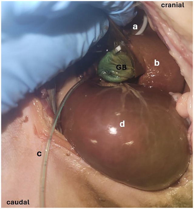
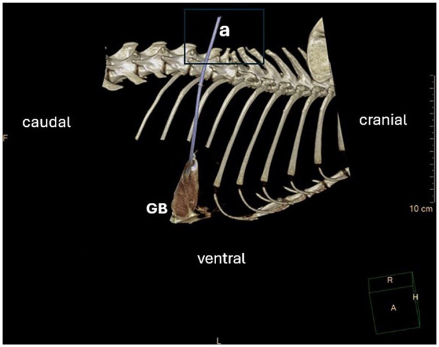
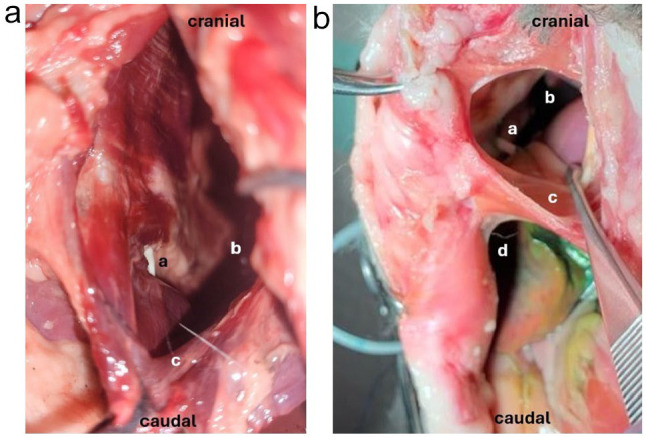
ObjectivesThe aim of the present study was to evaluate the feasibility and safety of percutaneous ultrasound- and fluoroscopy-guided cholecystostomy drain placement.MethodsAn experimental cadaveric study was conducted on 16 cat cadavers weighing between 2.5 and 6.4 kg. Two drain systems were tested for percutaneous ultrasound- and fluoroscopy-guided placement: the nephrostomy component of a subcutaneous urethral bypass system (SUB-nephrostomy drain) and a paediatric percutaneous access set (paediatric-nephrostomy drain). Ultrasound-guided cholecystocentesis was performed via the 8th-12th intercostal space. Using a Seldinger technique, a guidewire was advanced into the gallbladder over which the drains were then passed under fluoroscopic control. Protocol modification was required mid experiment. Gallbladders were filled via catheterisation of the common bile duct before cholecystocentesis. After fluoroscopy-confirmed cholecystostomy drain placement, CT scans were performed to assess drain position, iatrogenic organ injuries and leakage. Leak pressure testing was performed followed by anatomic dissection. Organ injuries were recorded and classified as minor, moderate or severe.ResultsSUB-nephrostomy drain placement was performed in 15 cats and placement into the gallbladder was feasible in two: one was passed before and one after technique modification. Paediatric-nephrostomy drain placement was tested in one cat. The gallbladder could not accommodate the drain size, placement was not feasible and the device was not further tested. A CT scan of the two cats with drain placement showed a moderate amount of free peritoneal contrast, no pleural space penetration and one liver injury. Leakage occurred at a pressure of 4.5 cm H2O. For all drains, injuries recorded during anatomic dissection were to the liver, pleural space and gallbladder. The majority of injuries were classified as minor.Conclusions and relevancePercutaneous placement of cholecystostomy drains was not feasible with the method and devices tested. Further studies are needed to investigate alternative techniques in cats.
期刊介绍:
JFMS is an international, peer-reviewed journal aimed at both practitioners and researchers with an interest in the clinical veterinary healthcare of domestic cats. The journal is published monthly in two formats: ‘Classic’ editions containing high-quality original papers on all aspects of feline medicine and surgery, including basic research relevant to clinical practice; and dedicated ‘Clinical Practice’ editions primarily containing opinionated review articles providing state-of-the-art information for feline clinicians, along with other relevant articles such as consensus guidelines.
 求助内容:
求助内容: 应助结果提醒方式:


