{"title":"Hepatic Angiosarcoma Presenting as Ascites: Case Report of a Rare yet Lethal Disease.","authors":"Archit Garg, Mehak Bassi, Capecomorin Pitchumoni, Arkady Broder","doi":"10.1159/000546375","DOIUrl":null,"url":null,"abstract":"<p><strong>Introduction: </strong>Angiosarcomas, constituting less than 1% of all sarcomas, are rare soft tissue tumors originating from the endothelial cells. Hepatic angiosarcoma (HAS) is a rare and aggressive primary hepatic malignancy accounting for only 0.5%-2% of all liver tumors. The patients often endorse nonspecific symptoms like vague abdominal pain, nausea, vomiting, and jaundice making the diagnosis challenging. Most patients succumb to death within 6 months of diagnosis due to liver failure or hemorrhage from spontaneous rupture of HAS. Therapeutic guidelines remain undefined, and management often involves a multidisciplinary approach. Surgical resection is the only potentially curative option, which has been shown to be most beneficial when HAS is limited to one lobe. Hepatic artery embolization is used in the case of rupture of HAS. Chemotherapy can be used for palliative care in cases of advanced tumors. We present a fatal case of metastatic HAS to underscore diagnostic pitfalls and therapeutic challenges.</p><p><strong>Case description: </strong>A 56-year-old male presented with 2 months of abdominal pain, distension, fatigue, and weight loss. Imaging revealed multifocal hypodense liver and splenic lesions. Laboratory findings included severe anemia (Hb 6.1 g/dL), thrombocytopenia (63 × 10<sup>3</sup>/mm<sup>3</sup>), and elevated liver enzymes. Ascitic fluid analysis demonstrated exudative, bloody ascites (SAAG <1.1) without malignant cytology. Liver biopsy confirmed HAS, showing atypical spindle cells infiltrating vascular channels, positive for CD34 and factor VIII. Despite transfusions, paracentesis, and palliative care, the patient developed disseminated intravascular coagulation and died 2 weeks post-diagnosis.</p><p><strong>Conclusion: </strong>HAS is a rapidly fatal malignancy often diagnosed at advanced stages due to nonspecific symptoms and lack of definitive risk factors in most cases. Multidisciplinary collaboration is essential for symptom management, though treatment options remain limited, and prognosis is poor. Therefore, it becomes imperative for clinicians to keep in mind the common presentation of a rare but lethal disease.</p>","PeriodicalId":9614,"journal":{"name":"Case Reports in Gastroenterology","volume":"19 1","pages":"488-495"},"PeriodicalIF":0.6000,"publicationDate":"2025-07-02","publicationTypes":"Journal Article","fieldsOfStudy":null,"isOpenAccess":false,"openAccessPdf":"https://www.ncbi.nlm.nih.gov/pmc/articles/PMC12215201/pdf/","citationCount":"0","resultStr":null,"platform":"Semanticscholar","paperid":null,"PeriodicalName":"Case Reports in Gastroenterology","FirstCategoryId":"1085","ListUrlMain":"https://doi.org/10.1159/000546375","RegionNum":0,"RegionCategory":null,"ArticlePicture":[],"TitleCN":null,"AbstractTextCN":null,"PMCID":null,"EPubDate":"2025/1/1 0:00:00","PubModel":"eCollection","JCR":"Q4","JCRName":"GASTROENTEROLOGY & HEPATOLOGY","Score":null,"Total":0}
引用次数: 0
Abstract
Introduction: Angiosarcomas, constituting less than 1% of all sarcomas, are rare soft tissue tumors originating from the endothelial cells. Hepatic angiosarcoma (HAS) is a rare and aggressive primary hepatic malignancy accounting for only 0.5%-2% of all liver tumors. The patients often endorse nonspecific symptoms like vague abdominal pain, nausea, vomiting, and jaundice making the diagnosis challenging. Most patients succumb to death within 6 months of diagnosis due to liver failure or hemorrhage from spontaneous rupture of HAS. Therapeutic guidelines remain undefined, and management often involves a multidisciplinary approach. Surgical resection is the only potentially curative option, which has been shown to be most beneficial when HAS is limited to one lobe. Hepatic artery embolization is used in the case of rupture of HAS. Chemotherapy can be used for palliative care in cases of advanced tumors. We present a fatal case of metastatic HAS to underscore diagnostic pitfalls and therapeutic challenges.
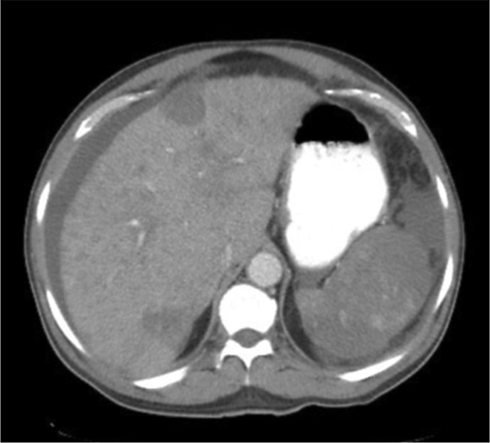
Case description: A 56-year-old male presented with 2 months of abdominal pain, distension, fatigue, and weight loss. Imaging revealed multifocal hypodense liver and splenic lesions. Laboratory findings included severe anemia (Hb 6.1 g/dL), thrombocytopenia (63 × 103/mm3), and elevated liver enzymes. Ascitic fluid analysis demonstrated exudative, bloody ascites (SAAG <1.1) without malignant cytology. Liver biopsy confirmed HAS, showing atypical spindle cells infiltrating vascular channels, positive for CD34 and factor VIII. Despite transfusions, paracentesis, and palliative care, the patient developed disseminated intravascular coagulation and died 2 weeks post-diagnosis.
Conclusion: HAS is a rapidly fatal malignancy often diagnosed at advanced stages due to nonspecific symptoms and lack of definitive risk factors in most cases. Multidisciplinary collaboration is essential for symptom management, though treatment options remain limited, and prognosis is poor. Therefore, it becomes imperative for clinicians to keep in mind the common presentation of a rare but lethal disease.
 求助内容:
求助内容: 应助结果提醒方式:


