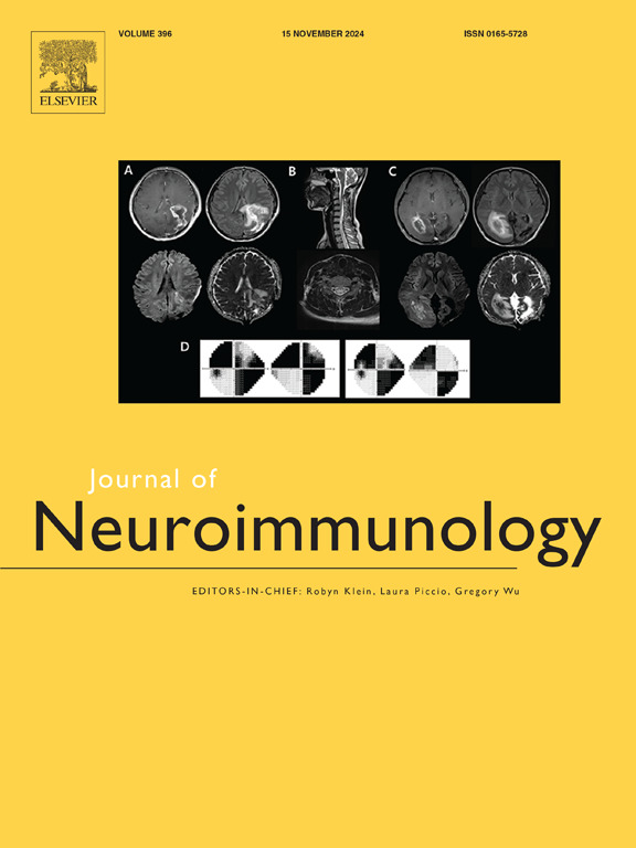
Association of ADHD symptoms, pain, and tics with anti-thalamus antibodies in cerebrospinal fluid
IF 2.5
4区 医学
Q3 IMMUNOLOGY
引用次数: 0
Abstract
Introduction
Complex mixed presentations of severe mental disorders (SMD) with treatment resistance pose major challenges in clinical practice. The role of novel neuronal antibodies in cerebrospinal fluid (CSF) is largely unexamined in this context.
Methods
A well-studied paradigmatic case of a 36-year-old female patient is reported.
Results
She presented with attention-deficit/hyperactivity disorder-like symptoms (including frequent sensory overload), severe pain (pre-diagnosed as fibromyalgia and somatoform pain disorder), and motor tics. In addition, she developed secondary depressive symptoms. Various psychopharmacological treatment attempts were unsuccessful or not tolerated. The diagnostic routine work-up with a wide range of blood tests, electroencephalography (EEG), routine magnetic resonance imaging (MRI), cerebrospinal fluid (CSF) analyses, and [18F]fluorodeoxyglucose positron emission tomography revealed no clear pathological findings. Tissue-based assays using CSF material found strong immunoglobulin G antibody staining specifically directed against a cell population in the thalamus. Neurotransmitter measurements detected low GABA, glutamate and serotonin concentrations as well as high dopamine levels in the CSF. Different MRI-based analyses indicated no neurostructural alterations in the thalamus; however, left mesiotemporal volume loss was identified. The independent component analysis of the EEG showed left temporal theta waves, partly resembling spike-wave-complexes. Immunotherapy using high-dose steroids resulted in a partial improvement with subjectively reduced stimulus overload, intermediate disappearance of pain, and fewer tics. The improvement could not be objectified psychometrically/neuropsychologically. The mesiotemporal volume loss was no longer present. There were no relevant changes in further research MRI measurements of the thalamus including arterial spin labeling, diffusion tensor imaging, and diffusion microstructure imaging from pre to post-immunotherapy.
Discussion
Novel antibodies against strategic brain structures, such as the thalamus, might be associated with some complex SMD. Further immunopsychiatric research in this direction holds promise for a better understanding of similar patients.
ADHD症状、疼痛和抽搐与脑脊液中抗丘脑抗体的关系
严重精神障碍(SMD)复杂的混合表现和治疗耐药性在临床实践中构成了重大挑战。在这种情况下,新型神经元抗体在脑脊液(CSF)中的作用在很大程度上尚未得到检验。方法报告一例36岁女性患者的典型病例。结果患者表现为注意缺陷/多动障碍样症状(包括频繁的感觉超负荷)、剧烈疼痛(预诊断为纤维肌痛和躯体形式疼痛障碍)和运动抽搐。此外,她还出现了继发性抑郁症状。各种心理药理学治疗尝试都不成功或不耐受。诊断常规检查包括广泛的血液检查、脑电图(EEG)、常规磁共振成像(MRI)、脑脊液(CSF)分析和[18F]氟脱氧葡萄糖正电子发射断层扫描未发现明确的病理发现。使用脑脊液材料进行的基于组织的检测发现,强烈的免疫球蛋白G抗体染色特异性地针对丘脑中的细胞群。神经递质测量检测到脑脊液中低GABA、谷氨酸和血清素浓度以及高多巴胺水平。不同的核磁共振分析表明,丘脑没有神经结构改变;然而,左颞叶体积损失被确定。脑电图独立分量分析显示左颞波,部分类似于尖波复合体。使用大剂量类固醇的免疫疗法导致部分改善,主观上减少了刺激过载,疼痛中度消失,抽搐减少。这种改善不能在心理测量学/神经心理学上客观化。中颞叶容积损失不再存在。在进一步的研究中,从免疫治疗前到后,丘脑的MRI测量包括动脉自旋标记、弥散张量成像和弥散微结构成像没有相关的变化。针对大脑战略结构(如丘脑)的新型抗体可能与一些复杂的SMD有关。在这个方向上进一步的免疫精神病学研究有望更好地了解类似的患者。
本文章由计算机程序翻译,如有差异,请以英文原文为准。
求助全文
约1分钟内获得全文
求助全文
来源期刊

Journal of neuroimmunology
医学-免疫学
CiteScore
6.10
自引率
3.00%
发文量
154
审稿时长
37 days
期刊介绍:
The Journal of Neuroimmunology affords a forum for the publication of works applying immunologic methodology to the furtherance of the neurological sciences. Studies on all branches of the neurosciences, particularly fundamental and applied neurobiology, neurology, neuropathology, neurochemistry, neurovirology, neuroendocrinology, neuromuscular research, neuropharmacology and psychology, which involve either immunologic methodology (e.g. immunocytochemistry) or fundamental immunology (e.g. antibody and lymphocyte assays), are considered for publication.
 求助内容:
求助内容: 应助结果提醒方式:


