Jae Seok Bae, Jeong Min Lee, Jeong Hee Yoon, Jae Hyun Kim, Sun Kyung Jeon, Jeongin Yoo
{"title":"Value of threshold growth for the diagnosis of hepatocellular carcinoma using LI-RADS.","authors":"Jae Seok Bae, Jeong Min Lee, Jeong Hee Yoon, Jae Hyun Kim, Sun Kyung Jeon, Jeongin Yoo","doi":"10.1186/s40644-025-00902-z","DOIUrl":null,"url":null,"abstract":"<p><strong>Background: </strong>The utility of threshold growth (TG) in hepatocellular carcinoma (HCC) imaging remains contentious across major guidelines. This study aimed to investigate the diagnostic implications of TG in HCC diagnosis using the criteria set by the Liver Imaging Reporting and Data System (LI-RADS).</p><p><strong>Methods: </strong>In this single-center retrospective study, three radiologists independently evaluated pre-transplantation hepatobiliary agent-enhanced MR images and prior CT/MR images using LI-RADS v2018 in consecutive patients who underwent liver transplantation between January 2010 and November 2022. TG was defined as a ≥ 50% size increase in ≤ 6 months. Explanted livers served as reference standards. Frequencies of TG between HCCs and non-HCCs were compared using Fisher's exact test, and interobserver agreement was assessed using Fleiss κ statistics. The diagnostic performance of LI-RADS category 5 in the diagnosis of HCC was assessed with and without considering TG as a major feature. McNemar tests were used to compare results.</p><p><strong>Results: </strong>The cohort included 158 patients (mean age, 59.1 ± 7.5 years; 130 males) with 280 observations (207 HCCs, 5 non-HCC malignancies, and 68 benign lesions). TG was identified in 44 (15.7%) observations. Interobserver agreement on TG was moderate (κ = 0.280). Incorporating TG as a major feature significantly enhanced the sensitivity of LI-RADS category 5 in diagnosing HCC (33.8% vs. 40.6%, p < 0.001) without compromising specificity (100.0% vs. 94.5%, p = 0.125).</p><p><strong>Conclusions: </strong>Incorporating TG as a major criterion in LI-RADS category 5 enhanced the diagnostic sensitivity for HCC in liver transplant candidates with minimal impact on specificity. However, TG demonstrated a variable interobserver agreement.</p><p><strong>Trial registration: </strong>Not applicable.</p>","PeriodicalId":9548,"journal":{"name":"Cancer Imaging","volume":"25 1","pages":"84"},"PeriodicalIF":3.5000,"publicationDate":"2025-07-01","publicationTypes":"Journal Article","fieldsOfStudy":null,"isOpenAccess":false,"openAccessPdf":"https://www.ncbi.nlm.nih.gov/pmc/articles/PMC12217194/pdf/","citationCount":"0","resultStr":null,"platform":"Semanticscholar","paperid":null,"PeriodicalName":"Cancer Imaging","FirstCategoryId":"3","ListUrlMain":"https://doi.org/10.1186/s40644-025-00902-z","RegionNum":2,"RegionCategory":"医学","ArticlePicture":[],"TitleCN":null,"AbstractTextCN":null,"PMCID":null,"EPubDate":"","PubModel":"","JCR":"Q2","JCRName":"ONCOLOGY","Score":null,"Total":0}
引用次数: 0
Abstract
Background: The utility of threshold growth (TG) in hepatocellular carcinoma (HCC) imaging remains contentious across major guidelines. This study aimed to investigate the diagnostic implications of TG in HCC diagnosis using the criteria set by the Liver Imaging Reporting and Data System (LI-RADS).
Methods: In this single-center retrospective study, three radiologists independently evaluated pre-transplantation hepatobiliary agent-enhanced MR images and prior CT/MR images using LI-RADS v2018 in consecutive patients who underwent liver transplantation between January 2010 and November 2022. TG was defined as a ≥ 50% size increase in ≤ 6 months. Explanted livers served as reference standards. Frequencies of TG between HCCs and non-HCCs were compared using Fisher's exact test, and interobserver agreement was assessed using Fleiss κ statistics. The diagnostic performance of LI-RADS category 5 in the diagnosis of HCC was assessed with and without considering TG as a major feature. McNemar tests were used to compare results.
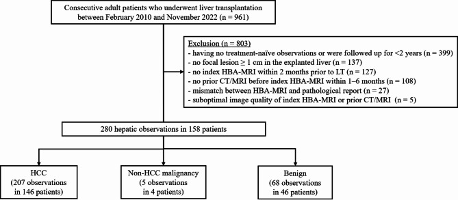
Results: The cohort included 158 patients (mean age, 59.1 ± 7.5 years; 130 males) with 280 observations (207 HCCs, 5 non-HCC malignancies, and 68 benign lesions). TG was identified in 44 (15.7%) observations. Interobserver agreement on TG was moderate (κ = 0.280). Incorporating TG as a major feature significantly enhanced the sensitivity of LI-RADS category 5 in diagnosing HCC (33.8% vs. 40.6%, p < 0.001) without compromising specificity (100.0% vs. 94.5%, p = 0.125).
Conclusions: Incorporating TG as a major criterion in LI-RADS category 5 enhanced the diagnostic sensitivity for HCC in liver transplant candidates with minimal impact on specificity. However, TG demonstrated a variable interobserver agreement.
Cancer ImagingONCOLOGY-RADIOLOGY, NUCLEAR MEDICINE & MEDICAL IMAGING
CiteScore
7.00
自引率
0.00%
发文量
66
审稿时长
>12 weeks
期刊介绍:
Cancer Imaging is an open access, peer-reviewed journal publishing original articles, reviews and editorials written by expert international radiologists working in oncology.
The journal encompasses CT, MR, PET, ultrasound, radionuclide and multimodal imaging in all kinds of malignant tumours, plus new developments, techniques and innovations. Topics of interest include:
Breast Imaging
Chest
Complications of treatment
Ear, Nose & Throat
Gastrointestinal
Hepatobiliary & Pancreatic
Imaging biomarkers
Interventional
Lymphoma
Measurement of tumour response
Molecular functional imaging
Musculoskeletal
Neuro oncology
Nuclear Medicine
Paediatric.

 求助内容:
求助内容: 应助结果提醒方式:


