Stephan Raad, Ali Al-Fatlawi, C Louise Wise, Christian Fottner, Simin Schadmand-Fischer, Mathias Schreckenberger, Matthias M Weber, Thomas J Musholt, Michael Schroeder, Matthias Miederer
{"title":"Imaging-derived biomarkers from <sup>68</sup>Ga-DOTATOC PET/CT scans to predict survival of patients with neuroendocrine tumors after PRRT with <sup>177</sup>Lu-DOTATATE.","authors":"Stephan Raad, Ali Al-Fatlawi, C Louise Wise, Christian Fottner, Simin Schadmand-Fischer, Mathias Schreckenberger, Matthias M Weber, Thomas J Musholt, Michael Schroeder, Matthias Miederer","doi":"10.1186/s40644-025-00899-5","DOIUrl":null,"url":null,"abstract":"<p><strong>Background: </strong>Neuroendocrine tumors have increased in prevalence and diversity in recent years and are often diagnosed at metastatic stages. Compared with nonradioactive systemic treatment with somatostatin analogs, peptide receptor radionuclide therapy (PRRT) has shown superior overall survival benefits for well-differentiated neuroendocrine tumor patients. This study aimed to identify biomarkers from <sup>68</sup>Ga‒DOTATOC PET/CT scans to predict survival in patients treated with PRRT in the clinic.</p><p><strong>Methodology: </strong>This retrospective study analyzed <sup>68</sup>Ga-DOTATOC PET/CT data from 67 NET patients undergoing PRRT. Tumor volumes and SUV metrics were segmented using standardized protocols. Radiomics features from liver metastases were extracted and preprocessed for analysis. Data were analysed via Kaplan-Meier, Cox regression, and PCA to evaluate the prognostic value of volumetric-, radiomics-, and clinicopathological parameters.</p><p><strong>Results: </strong>This study included scans from 67 patients with an average age of 67 years. The mean survival time was 46.5 months, with 43% of patients alive or lost to follow-up at the conclusion of data collection. Despite comprehensive analyses, neither volumetric parameters, including total tumor volume and organ-specific tumor volume, nor SUV values (SUVmax and SUVmean) were robust predictors of overall survival. K‒M and Cox regression analyses revealed no significant differences in survival between the high- and low-risk groups for these parameters. Furthermore, radiomics features extracted from liver metastases did not demonstrate significant prognostic value.</p><p><strong>Conclusion: </strong>Quantification of <sup>68</sup>Ga-DOTATOC PET/CT-derived parameters offers limited prognostic value for OS in NET patients who are receiving PRRT in clinical practice. These findings might emphasize the current robust integration of imaging in clinical decision-making for NET management.</p>","PeriodicalId":9548,"journal":{"name":"Cancer Imaging","volume":"25 1","pages":"81"},"PeriodicalIF":3.5000,"publicationDate":"2025-07-01","publicationTypes":"Journal Article","fieldsOfStudy":null,"isOpenAccess":false,"openAccessPdf":"https://www.ncbi.nlm.nih.gov/pmc/articles/PMC12211666/pdf/","citationCount":"0","resultStr":null,"platform":"Semanticscholar","paperid":null,"PeriodicalName":"Cancer Imaging","FirstCategoryId":"3","ListUrlMain":"https://doi.org/10.1186/s40644-025-00899-5","RegionNum":2,"RegionCategory":"医学","ArticlePicture":[],"TitleCN":null,"AbstractTextCN":null,"PMCID":null,"EPubDate":"","PubModel":"","JCR":"Q2","JCRName":"ONCOLOGY","Score":null,"Total":0}
引用次数: 0
Abstract
Background: Neuroendocrine tumors have increased in prevalence and diversity in recent years and are often diagnosed at metastatic stages. Compared with nonradioactive systemic treatment with somatostatin analogs, peptide receptor radionuclide therapy (PRRT) has shown superior overall survival benefits for well-differentiated neuroendocrine tumor patients. This study aimed to identify biomarkers from 68Ga‒DOTATOC PET/CT scans to predict survival in patients treated with PRRT in the clinic.
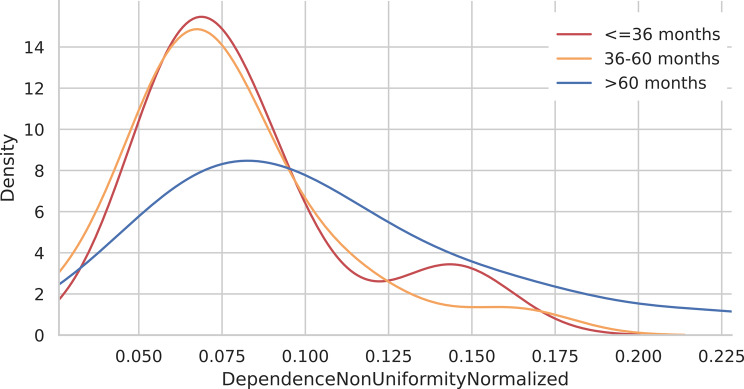
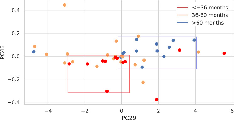
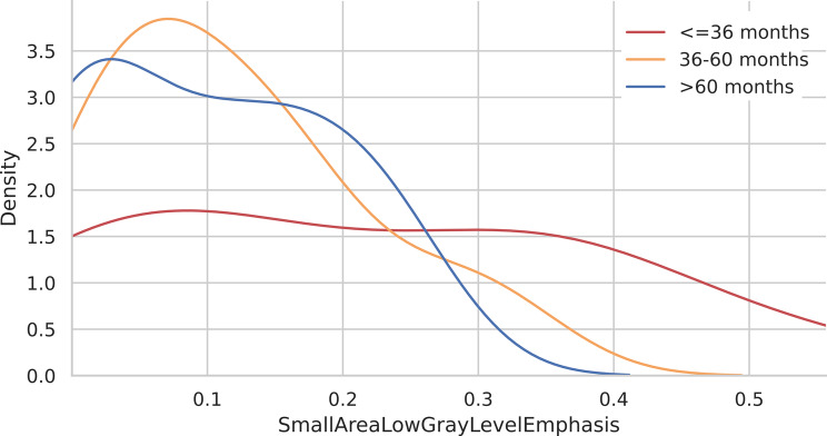
Methodology: This retrospective study analyzed 68Ga-DOTATOC PET/CT data from 67 NET patients undergoing PRRT. Tumor volumes and SUV metrics were segmented using standardized protocols. Radiomics features from liver metastases were extracted and preprocessed for analysis. Data were analysed via Kaplan-Meier, Cox regression, and PCA to evaluate the prognostic value of volumetric-, radiomics-, and clinicopathological parameters.
Results: This study included scans from 67 patients with an average age of 67 years. The mean survival time was 46.5 months, with 43% of patients alive or lost to follow-up at the conclusion of data collection. Despite comprehensive analyses, neither volumetric parameters, including total tumor volume and organ-specific tumor volume, nor SUV values (SUVmax and SUVmean) were robust predictors of overall survival. K‒M and Cox regression analyses revealed no significant differences in survival between the high- and low-risk groups for these parameters. Furthermore, radiomics features extracted from liver metastases did not demonstrate significant prognostic value.
Conclusion: Quantification of 68Ga-DOTATOC PET/CT-derived parameters offers limited prognostic value for OS in NET patients who are receiving PRRT in clinical practice. These findings might emphasize the current robust integration of imaging in clinical decision-making for NET management.
Cancer ImagingONCOLOGY-RADIOLOGY, NUCLEAR MEDICINE & MEDICAL IMAGING
CiteScore
7.00
自引率
0.00%
发文量
66
审稿时长
>12 weeks
期刊介绍:
Cancer Imaging is an open access, peer-reviewed journal publishing original articles, reviews and editorials written by expert international radiologists working in oncology.
The journal encompasses CT, MR, PET, ultrasound, radionuclide and multimodal imaging in all kinds of malignant tumours, plus new developments, techniques and innovations. Topics of interest include:
Breast Imaging
Chest
Complications of treatment
Ear, Nose & Throat
Gastrointestinal
Hepatobiliary & Pancreatic
Imaging biomarkers
Interventional
Lymphoma
Measurement of tumour response
Molecular functional imaging
Musculoskeletal
Neuro oncology
Nuclear Medicine
Paediatric.

 求助内容:
求助内容: 应助结果提醒方式:


