Viren Vasandani, Sean O'Leary, Ronak Gandhi, Elena Diller, Giri Movva, John Broussard, Vijaya Murthy
{"title":"Unraveling cerebrovascular involvement in EGPA through digital subtraction angiography: case presentation and systematic literature review.","authors":"Viren Vasandani, Sean O'Leary, Ronak Gandhi, Elena Diller, Giri Movva, John Broussard, Vijaya Murthy","doi":"10.1186/s41927-025-00518-7","DOIUrl":null,"url":null,"abstract":"<p><strong>Objectives: </strong>Eosinophilic granulomatosis with polyangiitis (EGPA) involves systemic inflammation of small to medium vessels, with central nervous system (CNS) involvement being rare. While CT (computed tomography) and MRI (magnetic resonance imaging) are standard for diagnosing CNS involvement, digital subtraction angiography (DSA) is infrequently used. We present a unique EGPA case with CNS involvement and review EGPA CNS vascular variations.</p><p><strong>Methods: </strong>We present a case of EGPA with CNS involvement, alongside a systematic review of the literature following PRISMA guidelines, querying three databases (PubMed/MEDLINE, SCOPUS, and Science Direct) up to September 2023 for case reports and series on EGPA with CNS involvement.</p><p><strong>Results: </strong>A 43-year-old presented with wheezing, multifocal neuropathy, leukocytosis, eosinophilia, positive ANA, and elevated CRP. Imaging revealed lung abnormalities. CT and MRI showed cerebral infarcts. CTA was negative, whereas DSA revealed bilateral segmental narrowing of anterior cerebral artery (ACA) branches and middle cerebral artery (MCA) branches. EGPA was confirmed, and treatment with steroids, cyclophosphamide, and azathioprine, led to remission. A systematic literature review of 27 EGPA cases with CNS involvement found a mean age 54.22 years, with common symptoms including extremity weakness (n = 8) and paresthesia (n = 5). Imaging techniques included MRI (n = 21), CT (n = 11), angiogram (n = 8), MRA (n = 4), CTA (n = 4), and MRV (n = 2), revealing stenosis of the bilateral ACA, vertebral artery, MCA, and basilar artery.</p><p><strong>Conclusion: </strong>Our findings suggest a potentially novel role for angiographic imaging in the comprehensive assessment of cerebrovascular involvement in EGPA.</p>","PeriodicalId":9150,"journal":{"name":"BMC Rheumatology","volume":"9 1","pages":"80"},"PeriodicalIF":2.5000,"publicationDate":"2025-07-01","publicationTypes":"Journal Article","fieldsOfStudy":null,"isOpenAccess":false,"openAccessPdf":"https://www.ncbi.nlm.nih.gov/pmc/articles/PMC12219991/pdf/","citationCount":"0","resultStr":null,"platform":"Semanticscholar","paperid":null,"PeriodicalName":"BMC Rheumatology","FirstCategoryId":"1085","ListUrlMain":"https://doi.org/10.1186/s41927-025-00518-7","RegionNum":0,"RegionCategory":null,"ArticlePicture":[],"TitleCN":null,"AbstractTextCN":null,"PMCID":null,"EPubDate":"","PubModel":"","JCR":"Q3","JCRName":"RHEUMATOLOGY","Score":null,"Total":0}
引用次数: 0
Abstract
Objectives: Eosinophilic granulomatosis with polyangiitis (EGPA) involves systemic inflammation of small to medium vessels, with central nervous system (CNS) involvement being rare. While CT (computed tomography) and MRI (magnetic resonance imaging) are standard for diagnosing CNS involvement, digital subtraction angiography (DSA) is infrequently used. We present a unique EGPA case with CNS involvement and review EGPA CNS vascular variations.
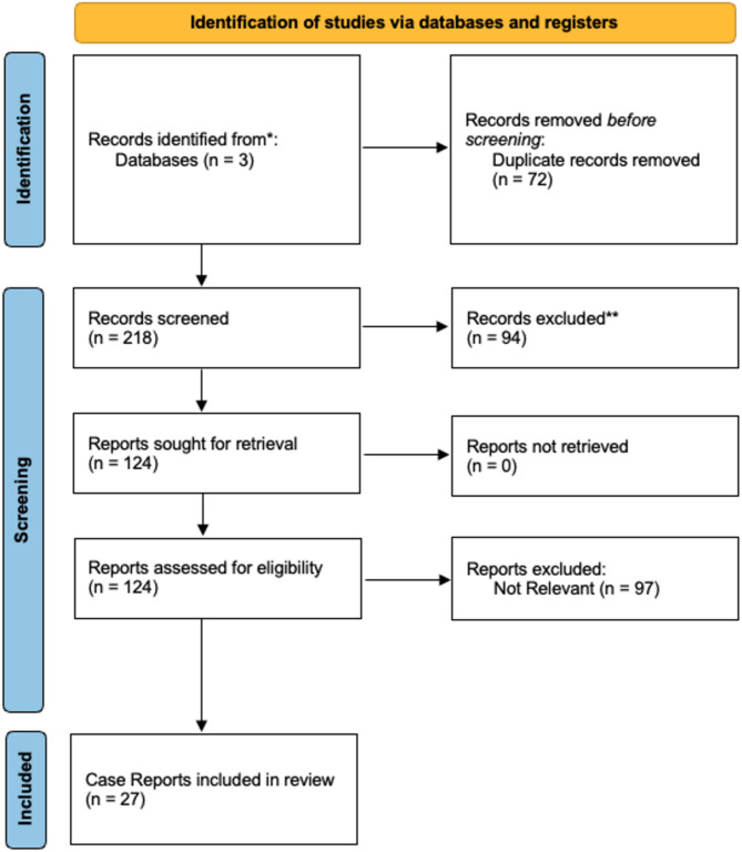
Methods: We present a case of EGPA with CNS involvement, alongside a systematic review of the literature following PRISMA guidelines, querying three databases (PubMed/MEDLINE, SCOPUS, and Science Direct) up to September 2023 for case reports and series on EGPA with CNS involvement.
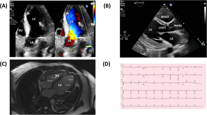
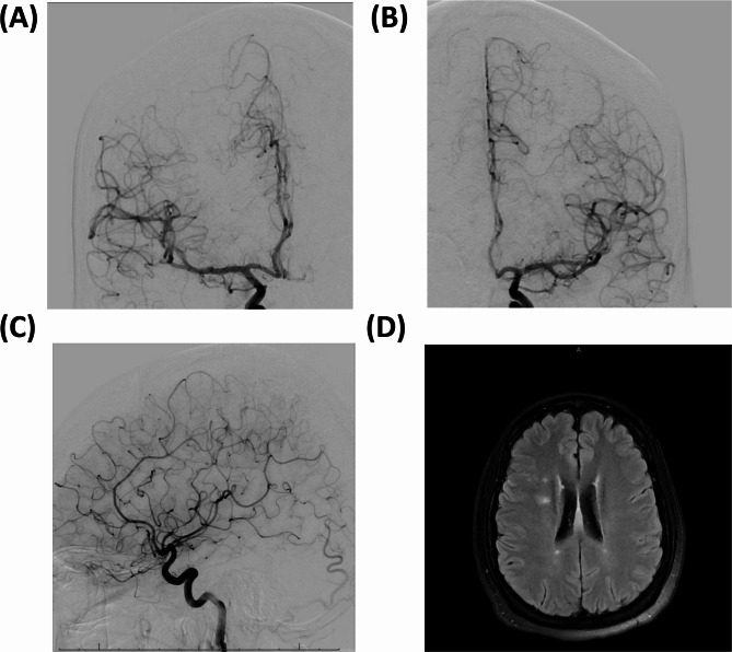
Results: A 43-year-old presented with wheezing, multifocal neuropathy, leukocytosis, eosinophilia, positive ANA, and elevated CRP. Imaging revealed lung abnormalities. CT and MRI showed cerebral infarcts. CTA was negative, whereas DSA revealed bilateral segmental narrowing of anterior cerebral artery (ACA) branches and middle cerebral artery (MCA) branches. EGPA was confirmed, and treatment with steroids, cyclophosphamide, and azathioprine, led to remission. A systematic literature review of 27 EGPA cases with CNS involvement found a mean age 54.22 years, with common symptoms including extremity weakness (n = 8) and paresthesia (n = 5). Imaging techniques included MRI (n = 21), CT (n = 11), angiogram (n = 8), MRA (n = 4), CTA (n = 4), and MRV (n = 2), revealing stenosis of the bilateral ACA, vertebral artery, MCA, and basilar artery.
Conclusion: Our findings suggest a potentially novel role for angiographic imaging in the comprehensive assessment of cerebrovascular involvement in EGPA.

 求助内容:
求助内容: 应助结果提醒方式:


