Hadi Tavakolikazerooni, Hao Yu, Saif Ullah, Wael Ennab, Dagan Mao
{"title":"Melatonin mitigates PGF-induced apoptosis during luteal regression in heat-exposed rats.","authors":"Hadi Tavakolikazerooni, Hao Yu, Saif Ullah, Wael Ennab, Dagan Mao","doi":"10.1590/1984-3143-AR2024-0122","DOIUrl":null,"url":null,"abstract":"<p><p>This study investigates the protective effects of melatonin against heat exposure during PGF-induced luteal regression in rats. Seventy-five PMSG and hCG primed rats were divided into three groups: non-heat-exposure (NHE), heat-exposure (HE), and melatonin <i>plus</i> heat-exposure (M+HE). The HE group underwent daily heat exposure (41°C for 2 h) for 7 days, while the M+HE group received intraperitoneal injection of melatonin (10 mg/kg body weight) before each heat session. On Day 7, PGF was administered, and ovarian samples were collected at 0, 1, 2, 8, and 24 h post-PGF. One set of ovaries was processed for histological analysis, including H&E staining, immunohistochemistry, and transmission electron microscopy and the other set was processed for Western blot for apoptotic protein expression. Results showed that heat exposure increased ovarian weight, disrupted follicular development, and elevated ovarian apoptotic markers (Caspase-3 and Bax), leading to luteal cell damage. Melatonin preserved ovarian weight, improved follicular and luteal structure, reduced atretic follicles, and mitigated luteal cell degeneration. In addition, melatonin decreased apoptotic marker expression and the Bax/Bcl-2 ratio, particularly at 16 and 24 h. These findings suggest melatonin protects luteal cells from heat-induced apoptosis during PGF-triggered regression, supporting reproductive function.</p>","PeriodicalId":7889,"journal":{"name":"Animal Reproduction","volume":"22 2","pages":"e20240122"},"PeriodicalIF":2.1000,"publicationDate":"2025-06-30","publicationTypes":"Journal Article","fieldsOfStudy":null,"isOpenAccess":false,"openAccessPdf":"https://www.ncbi.nlm.nih.gov/pmc/articles/PMC12212464/pdf/","citationCount":"0","resultStr":null,"platform":"Semanticscholar","paperid":null,"PeriodicalName":"Animal Reproduction","FirstCategoryId":"97","ListUrlMain":"https://doi.org/10.1590/1984-3143-AR2024-0122","RegionNum":4,"RegionCategory":"农林科学","ArticlePicture":[],"TitleCN":null,"AbstractTextCN":null,"PMCID":null,"EPubDate":"2025/1/1 0:00:00","PubModel":"eCollection","JCR":"Q2","JCRName":"AGRICULTURE, DAIRY & ANIMAL SCIENCE","Score":null,"Total":0}
引用次数: 0
Abstract
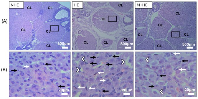
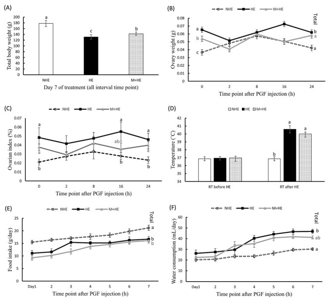
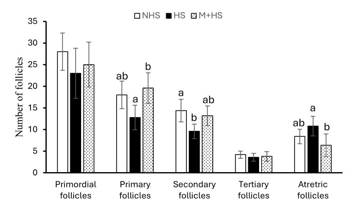
This study investigates the protective effects of melatonin against heat exposure during PGF-induced luteal regression in rats. Seventy-five PMSG and hCG primed rats were divided into three groups: non-heat-exposure (NHE), heat-exposure (HE), and melatonin plus heat-exposure (M+HE). The HE group underwent daily heat exposure (41°C for 2 h) for 7 days, while the M+HE group received intraperitoneal injection of melatonin (10 mg/kg body weight) before each heat session. On Day 7, PGF was administered, and ovarian samples were collected at 0, 1, 2, 8, and 24 h post-PGF. One set of ovaries was processed for histological analysis, including H&E staining, immunohistochemistry, and transmission electron microscopy and the other set was processed for Western blot for apoptotic protein expression. Results showed that heat exposure increased ovarian weight, disrupted follicular development, and elevated ovarian apoptotic markers (Caspase-3 and Bax), leading to luteal cell damage. Melatonin preserved ovarian weight, improved follicular and luteal structure, reduced atretic follicles, and mitigated luteal cell degeneration. In addition, melatonin decreased apoptotic marker expression and the Bax/Bcl-2 ratio, particularly at 16 and 24 h. These findings suggest melatonin protects luteal cells from heat-induced apoptosis during PGF-triggered regression, supporting reproductive function.
期刊介绍:
Animal Reproduction (AR) publishes original scientific papers and invited literature reviews, in the form of Basic Research, Biotechnology, Applied Research and Review Articles, with the goal of contributing to a better understanding of phenomena related to animal reproduction.
The scope of the journal applies to students, researchers and practitioners in the fields of veterinary, biology and animal science, also being of interest to practitioners of human medicine. Animal Reproduction Journal is the official organ of the Brazilian College of Animal Reproduction in Brazil.
 求助内容:
求助内容: 应助结果提醒方式:


