{"title":"Transverse navicular bone fracture in a hindlimb of a young Warmblood mare","authors":"P. Tóth, A. Nagy","doi":"10.1111/eve.14129","DOIUrl":null,"url":null,"abstract":"<p>A 2.5-year-old Warmblood filly presented with an acute onset of severe right hindlimb lameness and pastern region swelling of 2 weeks' duration. Radiographic examination revealed a transverse (horizontal) displaced fracture of the navicular bone and subluxation of the distal interphalangeal (DIP) joint. Computed tomographic (CT) examination, including DIP joint contrast arthrography, identified a further fragment in the plantar pouch of the DIP joint and revealed no communication between the DIP joint and the navicular bursa. Six months later the horse showed only mild lameness at trot. Computed tomographic examination revealed a partial osseous union between the large proximal and distal fragments. Ten months following the injury the mare was pasture sound and in foal.</p>","PeriodicalId":11786,"journal":{"name":"Equine Veterinary Education","volume":"37 8","pages":"e134-e140"},"PeriodicalIF":0.8000,"publicationDate":"2025-02-24","publicationTypes":"Journal Article","fieldsOfStudy":null,"isOpenAccess":false,"openAccessPdf":"https://onlinelibrary.wiley.com/doi/epdf/10.1111/eve.14129","citationCount":"0","resultStr":null,"platform":"Semanticscholar","paperid":null,"PeriodicalName":"Equine Veterinary Education","FirstCategoryId":"97","ListUrlMain":"https://beva.onlinelibrary.wiley.com/doi/10.1111/eve.14129","RegionNum":4,"RegionCategory":"农林科学","ArticlePicture":[],"TitleCN":null,"AbstractTextCN":null,"PMCID":null,"EPubDate":"","PubModel":"","JCR":"Q3","JCRName":"VETERINARY SCIENCES","Score":null,"Total":0}
引用次数: 0
Abstract
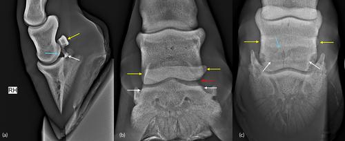
A 2.5-year-old Warmblood filly presented with an acute onset of severe right hindlimb lameness and pastern region swelling of 2 weeks' duration. Radiographic examination revealed a transverse (horizontal) displaced fracture of the navicular bone and subluxation of the distal interphalangeal (DIP) joint. Computed tomographic (CT) examination, including DIP joint contrast arthrography, identified a further fragment in the plantar pouch of the DIP joint and revealed no communication between the DIP joint and the navicular bursa. Six months later the horse showed only mild lameness at trot. Computed tomographic examination revealed a partial osseous union between the large proximal and distal fragments. Ten months following the injury the mare was pasture sound and in foal.
期刊介绍:
Equine Veterinary Education (EVE) is the official journal of post-graduate education of both the British Equine Veterinary Association (BEVA) and the American Association of Equine Practitioners (AAEP).
Equine Veterinary Education is a monthly, peer-reviewed, subscription-based journal, integrating clinical research papers, review articles and case reports from international sources, covering all aspects of medicine and surgery relating to equids. These papers facilitate the dissemination and implementation of new ideas and techniques relating to clinical veterinary practice, with the ultimate aim of promoting best practice. New developments are placed in perspective, encompassing new concepts and peer commentary. The target audience is veterinarians primarily engaged in the practise of equine medicine and surgery. The educational value of a submitted article is one of the most important criteria that are assessed when deciding whether to accept it for publication. Articles do not necessarily need to contain original or novel information but we welcome submission of this material. The educational value of an article may relate to articles published with it (e.g. a Case Report may not have direct educational value but an associated Clinical Commentary or Review Article published alongside it will enhance the educational value).


 求助内容:
求助内容: 应助结果提醒方式:


