Ultra-high-resolution and dual-energy computed tomography of carotid artery plaques differentiate symptomatic and asymptomatic patients by novel volumetric analysis.
Roland-Richard Macharzina, Simon Stemmler, Werner Vach, Thomas Winker, Jana Taron, Christopher L Schlett, Michael Weinbeck, Matthias Siepe, Martin Czerny, Fabian Bamberg, Thomas Zeller, Dirk Westermann, Martin Soschynski
{"title":"Ultra-high-resolution and dual-energy computed tomography of carotid artery plaques differentiate symptomatic and asymptomatic patients by novel volumetric analysis.","authors":"Roland-Richard Macharzina, Simon Stemmler, Werner Vach, Thomas Winker, Jana Taron, Christopher L Schlett, Michael Weinbeck, Matthias Siepe, Martin Czerny, Fabian Bamberg, Thomas Zeller, Dirk Westermann, Martin Soschynski","doi":"10.1093/icvts/ivaf158","DOIUrl":null,"url":null,"abstract":"<p><strong>Objectives: </strong>The indication for carotid endarterectomy (CEA) mainly relies on the degree of stenosis and neurological symptoms. Plaque vulnerability has been associated with stroke risk, but identification on single-energy computed tomography (CT) has yielded heterogeneous results and is not routinely applied to clinical diagnostics. Hence, we intended to analyse CEA specimens for vulnerability features using dual-source CT and correlate these features with the presence of preprocedural symptoms.</p><p><strong>Methods: </strong>CT was performed on 187 carotid plaque specimens using ultra-high-resolution and dual-energy imaging on a dual-source scanner. Plaques were separated into calcified versus non-calcified volumes and analysed concerning HU-density, calcifications and volumetric dual-energy indices (DEIs). Comparative statistical analysis of plaque characteristics was performed with respect to the presence of neurological symptoms.</p><p><strong>Results: </strong>The degree of stenosis of symptomatic and asymptomatic plaques was indifferent (69.2 ± 12.3% vs 66.3 ± 13.7%). The highest diagnostic accuracies were obtained by the % calcified volume (AUC 0.63 (0.54-0.71)), average whole plaque HU (AUC 0.71 (0.64-0.79)), profound calcification (AUC 0.74 (0.66-0.81)), calcification spots <1 mm (AUC 0.71 (0.63-0.79)) and spotty calcification (AUC 0.74 (0.66-0.82)). The diagnostic accuracy for symptomatic plaques was insignificant using average non-calcified plaque HU (AUC 0.59 (0.48-0.65)), but significant using average non-calcified plaque DEI (AUC 0.66 (0.58-0.74)).</p><p><strong>Conclusions: </strong>Symptomatic plaques were identified best by measuring density of the whole, calcified or non-calcified plaque and via spotty, profoundly localized and less dense calcification. A volumetric DEI identifies symptomatic plaques with non-calcified plaque characteristics more accurately than single-energy CT. Future clinical studies are necessary to confirm these findings in patients.</p>","PeriodicalId":73406,"journal":{"name":"Interdisciplinary cardiovascular and thoracic surgery","volume":" ","pages":""},"PeriodicalIF":0.0000,"publicationDate":"2025-07-03","publicationTypes":"Journal Article","fieldsOfStudy":null,"isOpenAccess":false,"openAccessPdf":"https://www.ncbi.nlm.nih.gov/pmc/articles/PMC12270255/pdf/","citationCount":"0","resultStr":null,"platform":"Semanticscholar","paperid":null,"PeriodicalName":"Interdisciplinary cardiovascular and thoracic surgery","FirstCategoryId":"1085","ListUrlMain":"https://doi.org/10.1093/icvts/ivaf158","RegionNum":0,"RegionCategory":null,"ArticlePicture":[],"TitleCN":null,"AbstractTextCN":null,"PMCID":null,"EPubDate":"","PubModel":"","JCR":"0","JCRName":"CARDIAC & CARDIOVASCULAR SYSTEMS","Score":null,"Total":0}
引用次数: 0
Abstract
Objectives: The indication for carotid endarterectomy (CEA) mainly relies on the degree of stenosis and neurological symptoms. Plaque vulnerability has been associated with stroke risk, but identification on single-energy computed tomography (CT) has yielded heterogeneous results and is not routinely applied to clinical diagnostics. Hence, we intended to analyse CEA specimens for vulnerability features using dual-source CT and correlate these features with the presence of preprocedural symptoms.
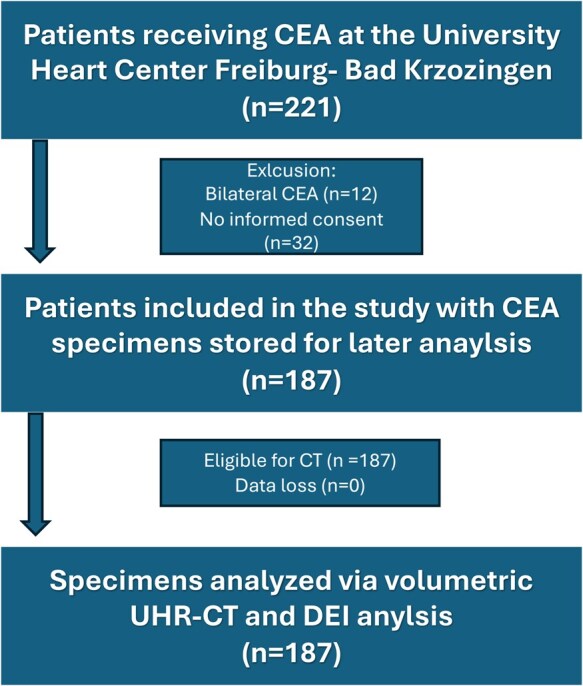
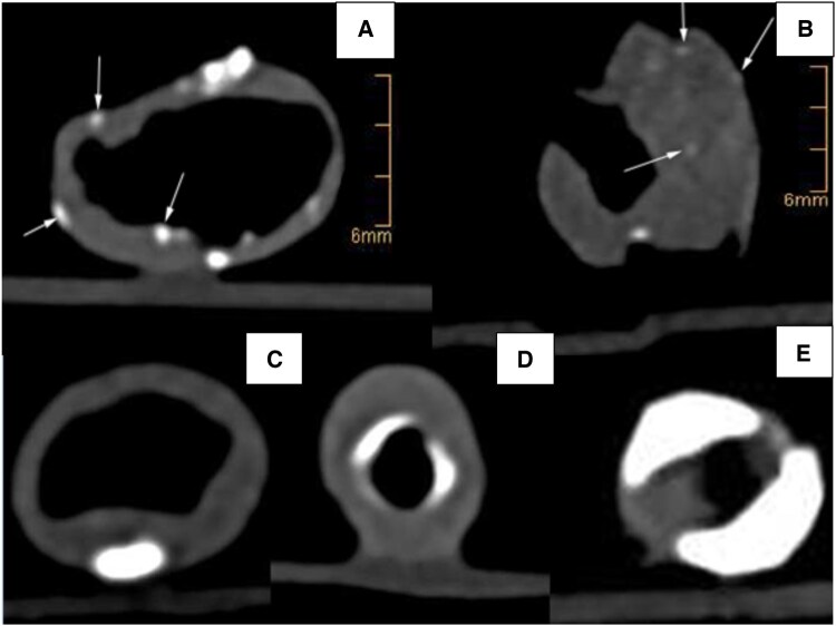
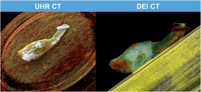
Methods: CT was performed on 187 carotid plaque specimens using ultra-high-resolution and dual-energy imaging on a dual-source scanner. Plaques were separated into calcified versus non-calcified volumes and analysed concerning HU-density, calcifications and volumetric dual-energy indices (DEIs). Comparative statistical analysis of plaque characteristics was performed with respect to the presence of neurological symptoms.
Results: The degree of stenosis of symptomatic and asymptomatic plaques was indifferent (69.2 ± 12.3% vs 66.3 ± 13.7%). The highest diagnostic accuracies were obtained by the % calcified volume (AUC 0.63 (0.54-0.71)), average whole plaque HU (AUC 0.71 (0.64-0.79)), profound calcification (AUC 0.74 (0.66-0.81)), calcification spots <1 mm (AUC 0.71 (0.63-0.79)) and spotty calcification (AUC 0.74 (0.66-0.82)). The diagnostic accuracy for symptomatic plaques was insignificant using average non-calcified plaque HU (AUC 0.59 (0.48-0.65)), but significant using average non-calcified plaque DEI (AUC 0.66 (0.58-0.74)).
Conclusions: Symptomatic plaques were identified best by measuring density of the whole, calcified or non-calcified plaque and via spotty, profoundly localized and less dense calcification. A volumetric DEI identifies symptomatic plaques with non-calcified plaque characteristics more accurately than single-energy CT. Future clinical studies are necessary to confirm these findings in patients.

 求助内容:
求助内容: 应助结果提醒方式:


