Jeong Heon Kim, Jeong Ho Woo, Minsu Kwon, Young Ho Jung, Seung-Ho Choi, Yoon Se Lee
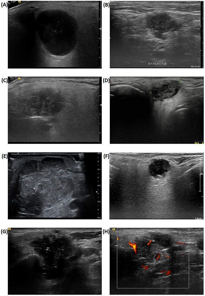
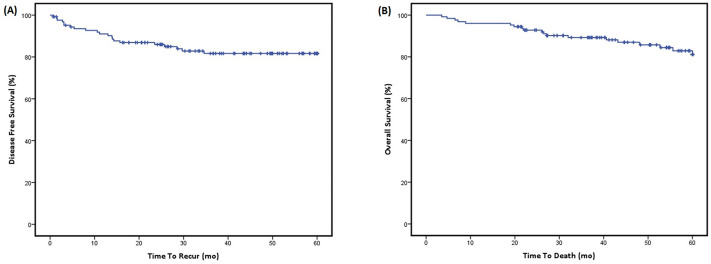
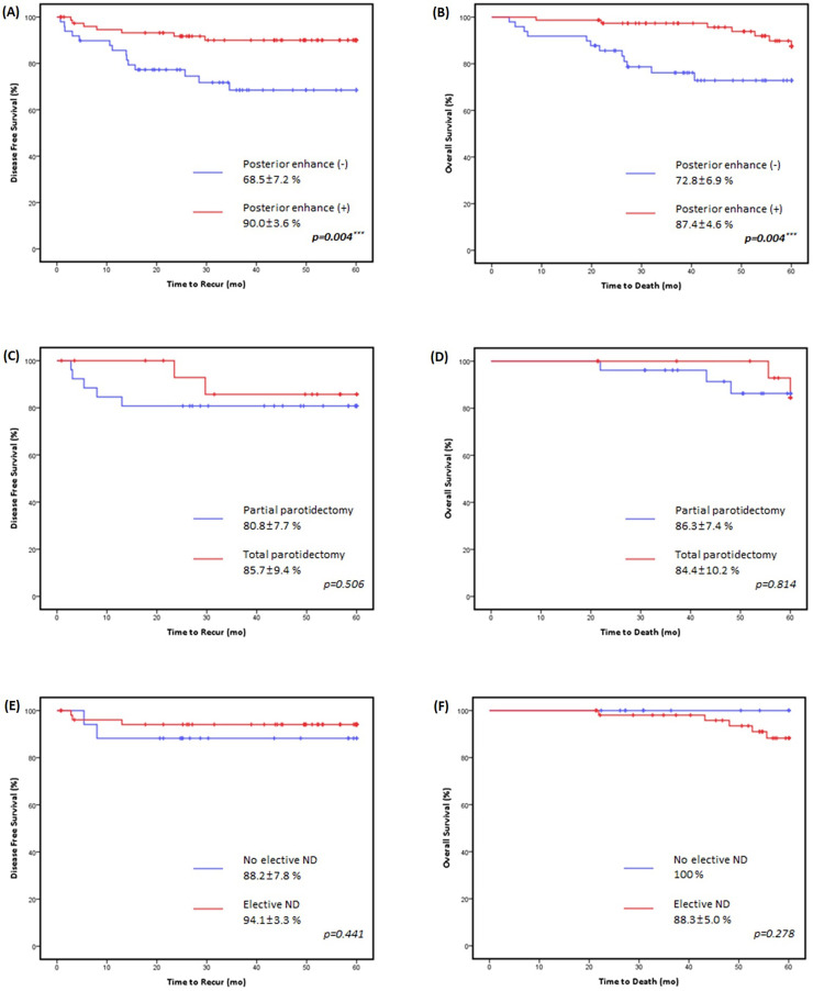
{"title":"Ultrasonographic findings as a prognostic factor in parotid cancer.","authors":"Jeong Heon Kim, Jeong Ho Woo, Minsu Kwon, Young Ho Jung, Seung-Ho Choi, Yoon Se Lee","doi":"10.1177/00368504251356179","DOIUrl":null,"url":null,"abstract":"<p><p>ObjectiveRecent advancements in high-resolution ultrasonography (US) have established it as a critical tool for evaluating parotid lesions. However, the lack of standardized diagnostic criteria limits the utility of US in determining malignancy. This study investigates the potential role of US as a prognostic factor in parotid cancer.MethodsPatients diagnosed with and surgically treated for parotid cancer at our tertiary referral center from January 2016 to December 2022 were included in this retrospective cohort study. We retrospectively obtained patient data including US images and clinical factors and analyzed their correlation with various adverse features and oncological outcomes, including five-year disease-free survival (5Y DFS) and overall survival (5Y OS).ResultsA total of 126 patients were included. The 5Y DFS and 5Y OS were 81.7% ± 3.7% and 81.2% ± 4.1% respectively. Multivariate analysis revealed that age (DFS; HR 2.75 [1.76-4.29], <i>p</i> = .023, OS; HR 3.38 [2.06-5.54], <i>p</i> = .014), clinical nodal stage (DFS; HR 5.87 [3.74-9.21], <i>p</i> < .001, OS; HR 9.34 [5.48-15.91], <i>p</i> < .001) and the presence of posterior enhancement artifact on US (DFS; HR 0.33 [0.21-0.53], <i>p</i> = .019, OS; HR 0.34 [0.20-0.57], <i>p</i> = .037) were significant variables. In patients with early-stage parotid cancer who showed posterior enhancement, the extent of surgery did not affect treatment outcomes.ConclusionPosterior acoustic enhancement on ultrasonography is a favorable prognostic factor in parotid cancer. For patients with early-stage cancer who demonstrate posterior enhancement, minimizing the extent of surgery does not compromise oncologic outcomes.</p>","PeriodicalId":56061,"journal":{"name":"Science Progress","volume":"108 3","pages":"368504251356179"},"PeriodicalIF":2.9000,"publicationDate":"2025-07-01","publicationTypes":"Journal Article","fieldsOfStudy":null,"isOpenAccess":false,"openAccessPdf":"https://www.ncbi.nlm.nih.gov/pmc/articles/PMC12217581/pdf/","citationCount":"0","resultStr":null,"platform":"Semanticscholar","paperid":null,"PeriodicalName":"Science Progress","FirstCategoryId":"103","ListUrlMain":"https://doi.org/10.1177/00368504251356179","RegionNum":4,"RegionCategory":"综合性期刊","ArticlePicture":[],"TitleCN":null,"AbstractTextCN":null,"PMCID":null,"EPubDate":"","PubModel":"","JCR":"Q2","JCRName":"MULTIDISCIPLINARY SCIENCES","Score":null,"Total":0}
引用次数: 0
Abstract
ObjectiveRecent advancements in high-resolution ultrasonography (US) have established it as a critical tool for evaluating parotid lesions. However, the lack of standardized diagnostic criteria limits the utility of US in determining malignancy. This study investigates the potential role of US as a prognostic factor in parotid cancer.MethodsPatients diagnosed with and surgically treated for parotid cancer at our tertiary referral center from January 2016 to December 2022 were included in this retrospective cohort study. We retrospectively obtained patient data including US images and clinical factors and analyzed their correlation with various adverse features and oncological outcomes, including five-year disease-free survival (5Y DFS) and overall survival (5Y OS).ResultsA total of 126 patients were included. The 5Y DFS and 5Y OS were 81.7% ± 3.7% and 81.2% ± 4.1% respectively. Multivariate analysis revealed that age (DFS; HR 2.75 [1.76-4.29], p = .023, OS; HR 3.38 [2.06-5.54], p = .014), clinical nodal stage (DFS; HR 5.87 [3.74-9.21], p < .001, OS; HR 9.34 [5.48-15.91], p < .001) and the presence of posterior enhancement artifact on US (DFS; HR 0.33 [0.21-0.53], p = .019, OS; HR 0.34 [0.20-0.57], p = .037) were significant variables. In patients with early-stage parotid cancer who showed posterior enhancement, the extent of surgery did not affect treatment outcomes.ConclusionPosterior acoustic enhancement on ultrasonography is a favorable prognostic factor in parotid cancer. For patients with early-stage cancer who demonstrate posterior enhancement, minimizing the extent of surgery does not compromise oncologic outcomes.
目的高分辨率超声检查(US)的最新进展使其成为评估腮腺病变的重要工具。然而,缺乏标准化的诊断标准限制了超声在确定恶性肿瘤中的应用。本研究探讨US作为腮腺癌预后因素的潜在作用。方法将2016年1月至2022年12月在我院三级转诊中心确诊并接受手术治疗的腮腺癌患者纳入回顾性队列研究。我们回顾性地获得了患者资料,包括US图像和临床因素,并分析了它们与各种不良特征和肿瘤预后的相关性,包括五年无病生存期(5Y DFS)和总生存期(5Y OS)。结果共纳入126例患者。5Y的DFS和OS分别为81.7%±3.7%和81.2%±4.1%。多变量分析显示,年龄(DFS;HR 2.75 [1.76-4.29], p =。023年,操作系统;HR 3.38 [2.06-5.54], p = 0.014),临床结期(DFS;HR 5.87 [3.74-9.21], p p p =。019年,操作系统;HR 0.34 [0.20-0.57], p = 0.037)为显著变量。在早期腮腺癌患者中,手术的程度对治疗结果没有影响。结论超声后路增强是腮腺癌预后的有利因素。对于表现出后部增强的早期癌症患者,最小化手术范围并不会影响肿瘤预后。
期刊介绍:
Science Progress has for over 100 years been a highly regarded review publication in science, technology and medicine. Its objective is to excite the readers'' interest in areas with which they may not be fully familiar but which could facilitate their interest, or even activity, in a cognate field.

 求助内容:
求助内容: 应助结果提醒方式:


