Nicole Bens, Arnold Chang, Richard Ortiz, Joshua Leaston, Praveen Kulkarni, Rosemarie Hightower, Sophia Prom, Nicholas O'Hare, Eno Ebong, Craig F Ferris
{"title":"Multimodal Magnetic Resonance Imaging with Mild Repetitive Head Injury in Awake Rats: Modeling the Human Experience and Clinical Condition.","authors":"Nicole Bens, Arnold Chang, Richard Ortiz, Joshua Leaston, Praveen Kulkarni, Rosemarie Hightower, Sophia Prom, Nicholas O'Hare, Eno Ebong, Craig F Ferris","doi":"10.1007/s12264-025-01438-9","DOIUrl":null,"url":null,"abstract":"<p><p>Mild repetitive head injury is a serious health problem with long-term negative consequences. Changes in brain neurobiology were assessed with MRI in a model of head injury designed to reflect the human experience. Rats were maintained on a reverse light-dark cycle and head impacted daily at 24 h intervals over three days while fully awake under red light illumination. There was no neuroradiological evidence of brain damage. Rats were imaged for changes in blood brain barrier permeability, edema and gray matter microarchitecture, and resting state functional connectivity. Data were registered to a 3D MRI rat atlas with 173 segmented brain areas providing site-specific information on each imaging modality. Changes in BBB permeability were minimal and localized to the hippocampus and cerebellum. There was evidence of cytotoxic edema in the basal ganglia, thalamus, and cerebellum. There was a global decrease in connectivity and an increase in gliosis in the thalamus, cerebellum, and hippocampus. This study shows a sequelae of neuropathology caused by mild repetitive head injury that is commonly observed in clinical practice using MRI in patients. As such, it may serve as a model for testing the efficacy of new therapeutics using any or all of the measures as biomarkers to assess drug efficacy.</p>","PeriodicalId":19314,"journal":{"name":"Neuroscience bulletin","volume":" ","pages":"1603-1616"},"PeriodicalIF":5.8000,"publicationDate":"2025-09-01","publicationTypes":"Journal Article","fieldsOfStudy":null,"isOpenAccess":false,"openAccessPdf":"https://www.ncbi.nlm.nih.gov/pmc/articles/PMC12433400/pdf/","citationCount":"0","resultStr":null,"platform":"Semanticscholar","paperid":null,"PeriodicalName":"Neuroscience bulletin","FirstCategoryId":"3","ListUrlMain":"https://doi.org/10.1007/s12264-025-01438-9","RegionNum":2,"RegionCategory":"医学","ArticlePicture":[],"TitleCN":null,"AbstractTextCN":null,"PMCID":null,"EPubDate":"2025/6/29 0:00:00","PubModel":"Epub","JCR":"Q1","JCRName":"NEUROSCIENCES","Score":null,"Total":0}
引用次数: 0
Abstract
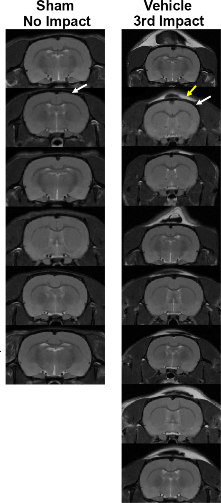
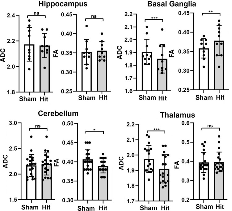
Mild repetitive head injury is a serious health problem with long-term negative consequences. Changes in brain neurobiology were assessed with MRI in a model of head injury designed to reflect the human experience. Rats were maintained on a reverse light-dark cycle and head impacted daily at 24 h intervals over three days while fully awake under red light illumination. There was no neuroradiological evidence of brain damage. Rats were imaged for changes in blood brain barrier permeability, edema and gray matter microarchitecture, and resting state functional connectivity. Data were registered to a 3D MRI rat atlas with 173 segmented brain areas providing site-specific information on each imaging modality. Changes in BBB permeability were minimal and localized to the hippocampus and cerebellum. There was evidence of cytotoxic edema in the basal ganglia, thalamus, and cerebellum. There was a global decrease in connectivity and an increase in gliosis in the thalamus, cerebellum, and hippocampus. This study shows a sequelae of neuropathology caused by mild repetitive head injury that is commonly observed in clinical practice using MRI in patients. As such, it may serve as a model for testing the efficacy of new therapeutics using any or all of the measures as biomarkers to assess drug efficacy.
期刊介绍:
Neuroscience Bulletin (NB), the official journal of the Chinese Neuroscience Society, is published monthly by Shanghai Institutes for Biological Sciences (SIBS), Chinese Academy of Sciences (CAS) and Springer.
NB aims to publish research advances in the field of neuroscience and promote exchange of scientific ideas within the community. The journal publishes original papers on various topics in neuroscience and focuses on potential disease implications on the nervous system. NB welcomes research contributions on molecular, cellular, or developmental neuroscience using multidisciplinary approaches and functional strategies. We feature full-length original articles, reviews, methods, letters to the editor, insights, and research highlights. As the official journal of the Chinese Neuroscience Society, which currently has more than 12,000 members in China, NB is devoted to facilitating communications between Chinese neuroscientists and their international colleagues. The journal is recognized as the most influential publication in neuroscience research in China.
 求助内容:
求助内容: 应助结果提醒方式:


