{"title":"Peroral Endoscopic Tumor Resection for Esophageal Leiomyoma with Accumulation of Fluorine-18-Fluorodeoxyglucose: A Case Report.","authors":"Takayuki Sawa, Osamu Dohi, Naoto Iwai, Katsuma Yamauchi, Mayuko Seya, Hajime Miyazaki, Hayato Fukui, Hiroaki Kitae, Tsugitaka Ishida, Yoshito Itoh","doi":"10.1159/000543564","DOIUrl":null,"url":null,"abstract":"<p><strong>Introduction: </strong>Esophageal leiomyomas are relatively common benign esophageal submucosal tumors (SMTs). Generally, benign tumors do not accumulate fluorine-18-fluorodeoxyglucose (FDG), but it is not rare for FDG to accumulate in uterine, duodenal, or esophageal leiomyomas. In our case, we performed peroral endoscopic tumor resection (POET) for an esophageal leiomyoma with FDG accumulation.</p><p><strong>Case presentation: </strong>A 40-year-old female with a history of surgery for right breast cancer underwent fluorine-18-fluorodeoxyglucose-positron emission tomography for surveillance examination and had no specific symptoms or notable clinical findings. A subepithelial tumor with intense FDG uptake (SUVmax, 5.49) was detected in the middle thoracic esophagus. The lesion appeared as a low-absorption area on contrast-enhanced CT and was confirmed to have an equivalent signal level as muscle tissue on MRI T2WI. Endoscopic examination revealed SMT 25 cm from the incisors. Endoscopic ultrasonography (EUS) revealed a 20 mm low-luminance mass, mainly located in the second and third layers. The histopathology diagnosis by EUS-fine-needle aspiration was leiomyoma. We decided to treat it with POET because malignancy could not be ruled out. The tumor was excised en bloc using POET without severe complications. The tumor diameter was 19 × 15 mm, and disordered spindle cells were observed. Desmin and αSMA were positive, and S100 protein was negative on immunohistochemical study. Therefore, the pathological diagnosis was a leiomyoma.</p><p><strong>Conclusion: </strong>In the present case, glucose transporter 1 expression was negative; however, we examined why the leiomyoma accumulated FDG. We suggest that awareness of leiomyoma with the accumulation of FDG exists in clinical practice.</p>","PeriodicalId":9614,"journal":{"name":"Case Reports in Gastroenterology","volume":"19 1","pages":"146-152"},"PeriodicalIF":0.6000,"publicationDate":"2025-03-12","publicationTypes":"Journal Article","fieldsOfStudy":null,"isOpenAccess":false,"openAccessPdf":"https://www.ncbi.nlm.nih.gov/pmc/articles/PMC11903047/pdf/","citationCount":"0","resultStr":null,"platform":"Semanticscholar","paperid":null,"PeriodicalName":"Case Reports in Gastroenterology","FirstCategoryId":"1085","ListUrlMain":"https://doi.org/10.1159/000543564","RegionNum":0,"RegionCategory":null,"ArticlePicture":[],"TitleCN":null,"AbstractTextCN":null,"PMCID":null,"EPubDate":"2025/1/1 0:00:00","PubModel":"eCollection","JCR":"Q4","JCRName":"GASTROENTEROLOGY & HEPATOLOGY","Score":null,"Total":0}
引用次数: 0
Abstract
Introduction: Esophageal leiomyomas are relatively common benign esophageal submucosal tumors (SMTs). Generally, benign tumors do not accumulate fluorine-18-fluorodeoxyglucose (FDG), but it is not rare for FDG to accumulate in uterine, duodenal, or esophageal leiomyomas. In our case, we performed peroral endoscopic tumor resection (POET) for an esophageal leiomyoma with FDG accumulation.
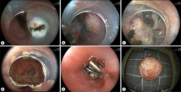
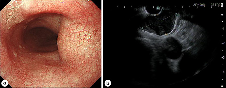
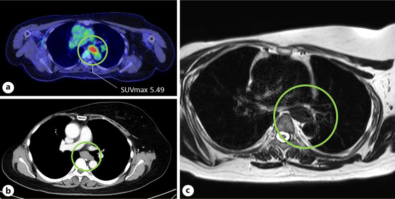
Case presentation: A 40-year-old female with a history of surgery for right breast cancer underwent fluorine-18-fluorodeoxyglucose-positron emission tomography for surveillance examination and had no specific symptoms or notable clinical findings. A subepithelial tumor with intense FDG uptake (SUVmax, 5.49) was detected in the middle thoracic esophagus. The lesion appeared as a low-absorption area on contrast-enhanced CT and was confirmed to have an equivalent signal level as muscle tissue on MRI T2WI. Endoscopic examination revealed SMT 25 cm from the incisors. Endoscopic ultrasonography (EUS) revealed a 20 mm low-luminance mass, mainly located in the second and third layers. The histopathology diagnosis by EUS-fine-needle aspiration was leiomyoma. We decided to treat it with POET because malignancy could not be ruled out. The tumor was excised en bloc using POET without severe complications. The tumor diameter was 19 × 15 mm, and disordered spindle cells were observed. Desmin and αSMA were positive, and S100 protein was negative on immunohistochemical study. Therefore, the pathological diagnosis was a leiomyoma.
Conclusion: In the present case, glucose transporter 1 expression was negative; however, we examined why the leiomyoma accumulated FDG. We suggest that awareness of leiomyoma with the accumulation of FDG exists in clinical practice.
 求助内容:
求助内容: 应助结果提醒方式:


