Mohammad Ghorbani, Ehsan Keykhosravi, Mohammad Hasanpour, Ali Abbasian Ardakani, Ehsan Mohammad Hosseini
{"title":"The Optimal Time for Postoperative Magnetic Resonance Imaging of the Sella in Patients With Pituitary Adenoma.","authors":"Mohammad Ghorbani, Ehsan Keykhosravi, Mohammad Hasanpour, Ali Abbasian Ardakani, Ehsan Mohammad Hosseini","doi":"10.32598/bcn.2023.5005.1","DOIUrl":null,"url":null,"abstract":"<p><strong>Introduction: </strong>Magnetic resonance imaging (MRI) is the preferred neuroradiologic tool for evaluating the sellar region. Pituitary adenomas account for about 15% of primary intracranial tumors. The optimal time for postoperative MRI of central nervous system neoplasms is 48 hours after surgery. Nevertheless, controversy exists regarding the timing of postoperative MRI in the sellar region. This study analyzed the sellar MRI findings of patients with pituitary adenoma at different times before and after surgery. Finally, we suggest the optimal time for postoperative sellar MRI imaging in patients with pituitary adenoma.</p><p><strong>Methods: </strong>A total of 28 patients with pituitary adenoma were evaluated. All patients did four sellar MRIs. The first MRI was done before surgery, and three were done 48 hours, two weeks, and three months after the surgery. Finally, the MRI findings at different times were compared to each other.</p><p><strong>Results: </strong>The pituitary gland and adenoma signals were constant at all time points. The signal of the packing material showed no differences in T1-weighted and T1-weighted with contrast sequences but showed changes in T2-weighted sequences.</p><p><strong>Conclusion: </strong>Contrary to other intracranial neoplasms, there were no apparent changes in MRI signal intensity during the 3 months after surgery in patients with pituitary adenoma. There was also no superiority of one time point for performing follow-up imaging.</p>","PeriodicalId":8701,"journal":{"name":"Basic and Clinical Neuroscience","volume":"15 5","pages":"649-658"},"PeriodicalIF":1.1000,"publicationDate":"2024-09-01","publicationTypes":"Journal Article","fieldsOfStudy":null,"isOpenAccess":false,"openAccessPdf":"https://www.ncbi.nlm.nih.gov/pmc/articles/PMC12198737/pdf/","citationCount":"0","resultStr":null,"platform":"Semanticscholar","paperid":null,"PeriodicalName":"Basic and Clinical Neuroscience","FirstCategoryId":"1085","ListUrlMain":"https://doi.org/10.32598/bcn.2023.5005.1","RegionNum":0,"RegionCategory":null,"ArticlePicture":[],"TitleCN":null,"AbstractTextCN":null,"PMCID":null,"EPubDate":"","PubModel":"","JCR":"Q4","JCRName":"NEUROSCIENCES","Score":null,"Total":0}
引用次数: 0
Abstract
Introduction: Magnetic resonance imaging (MRI) is the preferred neuroradiologic tool for evaluating the sellar region. Pituitary adenomas account for about 15% of primary intracranial tumors. The optimal time for postoperative MRI of central nervous system neoplasms is 48 hours after surgery. Nevertheless, controversy exists regarding the timing of postoperative MRI in the sellar region. This study analyzed the sellar MRI findings of patients with pituitary adenoma at different times before and after surgery. Finally, we suggest the optimal time for postoperative sellar MRI imaging in patients with pituitary adenoma.
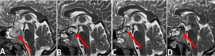
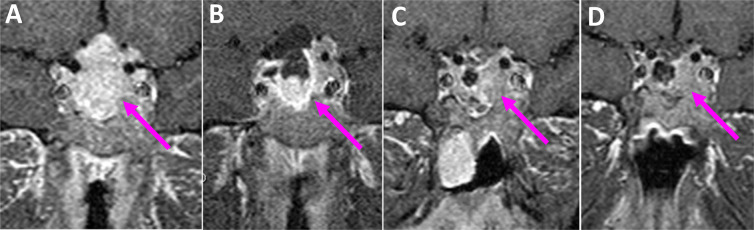
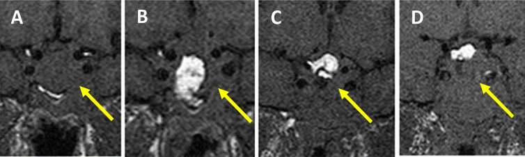
Methods: A total of 28 patients with pituitary adenoma were evaluated. All patients did four sellar MRIs. The first MRI was done before surgery, and three were done 48 hours, two weeks, and three months after the surgery. Finally, the MRI findings at different times were compared to each other.
Results: The pituitary gland and adenoma signals were constant at all time points. The signal of the packing material showed no differences in T1-weighted and T1-weighted with contrast sequences but showed changes in T2-weighted sequences.
Conclusion: Contrary to other intracranial neoplasms, there were no apparent changes in MRI signal intensity during the 3 months after surgery in patients with pituitary adenoma. There was also no superiority of one time point for performing follow-up imaging.
期刊介绍:
BCN is an international multidisciplinary journal that publishes editorials, original full-length research articles, short communications, reviews, methodological papers, commentaries, perspectives and “news and reports” in the broad fields of developmental, molecular, cellular, system, computational, behavioral, cognitive, and clinical neuroscience. No area in the neural related sciences is excluded from consideration, although priority is given to studies that provide applied insights into the functioning of the nervous system. BCN aims to advance our understanding of organization and function of the nervous system in health and disease, thereby improving the diagnosis and treatment of neural-related disorders. Manuscripts submitted to BCN should describe novel results generated by experiments that were guided by clearly defined aims or hypotheses. BCN aims to provide serious ties in interdisciplinary communication, accessibility to a broad readership inside Iran and the region and also in all other international academic sites, effective peer review process, and independence from all possible non-scientific interests. BCN also tries to empower national, regional and international collaborative networks in the field of neuroscience in Iran, Middle East, Central Asia and North Africa and to be the voice of the Iranian and regional neuroscience community in the world of neuroscientists. In this way, the journal encourages submission of editorials, review papers, commentaries, methodological notes and perspectives that address this scope.
 求助内容:
求助内容: 应助结果提醒方式:


