David L Chan, Alice Conner, Nick Pavlakis, Elizabeth Bailey, Alireza Aslani, Kathy Willowson, Connie Diakos, Elizabeth J Bernard, Stephen Clarke, Alexander Engel, Paul J Roach, Dale L Bailey
{"title":"[<sup>18</sup>F]FMISO PET in metastatic neuroendocrine tumours: a pilot study.","authors":"David L Chan, Alice Conner, Nick Pavlakis, Elizabeth Bailey, Alireza Aslani, Kathy Willowson, Connie Diakos, Elizabeth J Bernard, Stephen Clarke, Alexander Engel, Paul J Roach, Dale L Bailey","doi":"10.22038/aojnmb.2025.83664.1611","DOIUrl":null,"url":null,"abstract":"<p><strong>Objectives: </strong>The phenomenon of peripheral [<sup>68</sup>Ga]DOTATATE avidity without central avidity (which we have termed a \"DONUT\") has been observed in neuroendocrine neoplasm (NEN) lesions. There has been speculation as to whether this is due to hypoxia, de-differentiated disease or other causes. The presence of hypoxia may have prognostic and therapeutic implications, and was evaluated in these lesions using the PET hypoxia imaging biomarker [<sup>18</sup>F]FMISO.</p><p><strong>Methods: </strong>Prospective pilot study in patients with metastatic NENs with at least one DONUT lesion (central [<sup>68</sup>Ga]DOTATATE non-avidity). [<sup>18</sup>F]FDG and [<sup>18</sup>F]FMISO scans were acquired within 60 days of the [<sup>68</sup>Ga]DOTATATE PET/CT. [<sup>18</sup>F]FMISO scans were acquired as a dynamic scan over 20 mins from injection with a delayed image at 2 hours. The dynamic acquisition was analysed quantitatively using a graphical approach yielding parametric images of Influx Rate Constant and Volume of Distribution. [<sup>18</sup>F]FMISO uptake within the identified DONUT hole on the 2 hr delayed scan was qualitatively scored by two experienced nuclear medicine physicians as: 0 (no uptake), 1 (uptake less than normal liver), 2 (uptake equal to normal liver), or 3 (uptake greater than normal liver).</p><p><strong>Results: </strong>Ten patients were enrolled with primary sites including pancreas (n=3), small bowel (n=3), rectum (n=2), duodenum (n=1) and lung (n=1). Six subjects were scored 1, three subjects were scored 2, and one subject was scored 3. All lesions evaluated were located in the liver. Quantitative [<sup>18</sup>F]FMISO parametric imaging showed evidence of increased uptake rate (Ki) in the photopenic areas of the DONUT lesions in 8/10 subjects. Surrounding uptake rate in normal liver was extremely low. In the qualitative delayed image assessment, only one subject demonstrated [<sup>18</sup>F]FMISO uptake greater than surrounding normal liver (small bowel primary, G2).</p><p><strong>Conclusion: </strong>Only one of ten patients with DONUT lesions demonstrated increased [<sup>18</sup>F]FMISO uptake rate on delayed static imaging. In contrast, dynamic imaging demonstrated increased [<sup>18</sup>F]FMISO uptake rate in the region of [<sup>68</sup>Ga]DOTATATE photopenia on 8 of 10 patients. Future research using [<sup>18</sup>F]FMISO in NEN patients should incorporate dynamic imaging.</p>","PeriodicalId":8503,"journal":{"name":"Asia Oceania Journal of Nuclear Medicine and Biology","volume":"13 2","pages":"117-125"},"PeriodicalIF":0.0000,"publicationDate":"2025-01-01","publicationTypes":"Journal Article","fieldsOfStudy":null,"isOpenAccess":false,"openAccessPdf":"https://www.ncbi.nlm.nih.gov/pmc/articles/PMC12205124/pdf/","citationCount":"0","resultStr":null,"platform":"Semanticscholar","paperid":null,"PeriodicalName":"Asia Oceania Journal of Nuclear Medicine and Biology","FirstCategoryId":"1085","ListUrlMain":"https://doi.org/10.22038/aojnmb.2025.83664.1611","RegionNum":0,"RegionCategory":null,"ArticlePicture":[],"TitleCN":null,"AbstractTextCN":null,"PMCID":null,"EPubDate":"","PubModel":"","JCR":"Q3","JCRName":"Medicine","Score":null,"Total":0}
引用次数: 0
Abstract
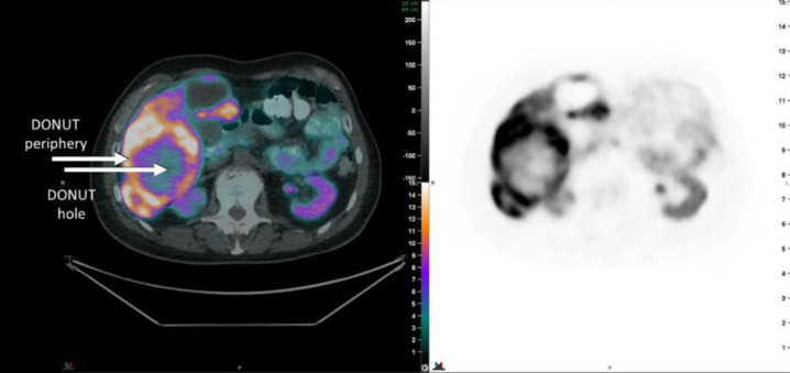
Objectives: The phenomenon of peripheral [68Ga]DOTATATE avidity without central avidity (which we have termed a "DONUT") has been observed in neuroendocrine neoplasm (NEN) lesions. There has been speculation as to whether this is due to hypoxia, de-differentiated disease or other causes. The presence of hypoxia may have prognostic and therapeutic implications, and was evaluated in these lesions using the PET hypoxia imaging biomarker [18F]FMISO.
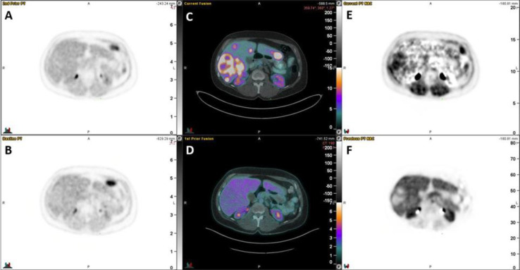
Methods: Prospective pilot study in patients with metastatic NENs with at least one DONUT lesion (central [68Ga]DOTATATE non-avidity). [18F]FDG and [18F]FMISO scans were acquired within 60 days of the [68Ga]DOTATATE PET/CT. [18F]FMISO scans were acquired as a dynamic scan over 20 mins from injection with a delayed image at 2 hours. The dynamic acquisition was analysed quantitatively using a graphical approach yielding parametric images of Influx Rate Constant and Volume of Distribution. [18F]FMISO uptake within the identified DONUT hole on the 2 hr delayed scan was qualitatively scored by two experienced nuclear medicine physicians as: 0 (no uptake), 1 (uptake less than normal liver), 2 (uptake equal to normal liver), or 3 (uptake greater than normal liver).
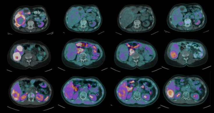
Results: Ten patients were enrolled with primary sites including pancreas (n=3), small bowel (n=3), rectum (n=2), duodenum (n=1) and lung (n=1). Six subjects were scored 1, three subjects were scored 2, and one subject was scored 3. All lesions evaluated were located in the liver. Quantitative [18F]FMISO parametric imaging showed evidence of increased uptake rate (Ki) in the photopenic areas of the DONUT lesions in 8/10 subjects. Surrounding uptake rate in normal liver was extremely low. In the qualitative delayed image assessment, only one subject demonstrated [18F]FMISO uptake greater than surrounding normal liver (small bowel primary, G2).
Conclusion: Only one of ten patients with DONUT lesions demonstrated increased [18F]FMISO uptake rate on delayed static imaging. In contrast, dynamic imaging demonstrated increased [18F]FMISO uptake rate in the region of [68Ga]DOTATATE photopenia on 8 of 10 patients. Future research using [18F]FMISO in NEN patients should incorporate dynamic imaging.

 求助内容:
求助内容: 应助结果提醒方式:


