Nanosensor for carcinoembryonic antigen (CEA) detection based on a selective growth-regulating mechanism
引用次数: 0
Abstract
Nanomaterial-based biosensors provide high sensitivity and versatility for the detection of target analytes. Particularly, gold nanomaterials are widely employed as signal transducers due to their unique plasmonic properties. When functionalized with aptamers, these nanostructures gain enhanced selectivity, rapid response capability, and cost-effectiveness. However, the instability of large gold nanoparticles can hinder the exploration of various shapes and sizes.
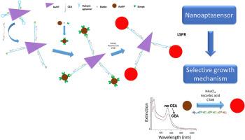
In this research, two different types of nanoparticles were used for the development of a carcinoembryonic antigen (CEA), a tumoral biomarker for colorectal cancer, nanosensor. It was proposed a system based on gold nanotriangles (AuNTs) functionalized with the amphiphilic polymer polyvinylpyrrolidone (AuNT@PVP) and a hairpin-shaped aptamer selective to CEA biotinylated at the 3′ end and thiolated at the 5′ end (AuNT@PVP@Apt). Specific recognition process was optimized expecting that CEA would trigger the unfolding of the hairpin structure, exposing biotin moieties that enabled subsequent binding of streptavidin-functionalized gold nanoparticles (AuNP@Strept, 3–5 nm in diameter). However, when analyzing the optical changes observed by UV–Vis–NIR spectroscopy and its correlation with the concentration of CEA, it was observed that the main mechanism involved in the detection was a selective growth mechanism of AuNPs, leading to a really simple and easy way of detecting CEA. The results obtained in this study highlight the intricacies of developing such nanosensors and introduce a novel detection method based on a selective growth mechanism that can be further used for detection.
基于选择性生长调节机制的癌胚抗原(CEA)检测纳米传感器
基于纳米材料的生物传感器为目标分析物的检测提供了高灵敏度和多功能性。特别是,金纳米材料由于其独特的等离子体特性而被广泛应用于信号换能器。当与适体功能化时,这些纳米结构获得了增强的选择性,快速反应能力和成本效益。然而,大型金纳米颗粒的不稳定性会阻碍对各种形状和尺寸的探索。在这项研究中,两种不同类型的纳米颗粒被用于发展癌胚抗原(CEA),结直肠癌的肿瘤生物标志物,纳米传感器。提出了一种基于两亲性聚合物聚乙烯吡咯烷酮(AuNT@PVP)功能化的金纳米三角形(AuNTs)和一个3 ‘端选择性CEA生物素化和5 ’端选择性硫代化的发夹形适体(AuNT@PVP@Apt)的体系。特异性识别过程被优化,期望CEA会触发发夹结构的展开,暴露生物素片段,使随后结合链霉亲和素功能化的金纳米颗粒(AuNP@Strept,直径3-5 nm)。然而,在分析UV-Vis-NIR光谱观察到的光学变化及其与CEA浓度的相关性时,发现检测的主要机制是AuNPs的选择性生长机制,这使得检测CEA的方法非常简单易行。本研究的结果强调了开发这种纳米传感器的复杂性,并介绍了一种基于选择性生长机制的新型检测方法,该方法可以进一步用于检测。
本文章由计算机程序翻译,如有差异,请以英文原文为准。
求助全文
约1分钟内获得全文
求助全文

 求助内容:
求助内容: 应助结果提醒方式:


