{"title":"Identifying the Primary Tumor Site and Distinguishing False-positives in Patients With Elevated Serum Carcinoembryonic Antigen.","authors":"Hiroaki Satoh, Takumi Sakamoto, Yutaka Takahashi, Kunihiko Miyazaki, Satoshi Inagawa","doi":"10.21873/invivo.14035","DOIUrl":null,"url":null,"abstract":"<p><strong>Background/aim: </strong>Carcinoembryonic antigen (CEA) is a tumor marker that is frequently evaluated clinically for gastrointestinal and lung cancers. CEA is a glycoprotein antigen, and only the \"value\" measured by enzyme-linked immunosorbent assay is provided in the clinical setting. At present, no method has been established to indicate whether the value is a false-positive elevation or whether there is a primary cancer site. To obtain clues on how to identify the originating site in patients with cancer with high CEA levels and to identify CEA false-positives in healthy individuals, we conducted an exploratory study.</p><p><strong>Patients and methods: </strong>A pilot study was performed using the multivariate analysis method and principal component analysis-discriminant analysis on proteomic results obtained using liquid chromatography-mass spectrometry (LC/MS) in two patients with lung cancer, one patient with gastric cancer, and one healthy control individual.</p><p><strong>Results: </strong>No differences in specific proteins associated with high CEA levels were detected between lung and gastric cancers using LC/MS. Therefore, we performed statistical analysis using principal component analysis-discriminant analysis to determine whether there were differences in the protein signal patterns obtained using LC/MS. The results showed that the plots obtained for each patient and the healthy control were located in different quadrants of the four-quadrant matrix scatter plot.</p><p><strong>Conclusion: </strong>Our results suggest the possibility of visually differentiating the primary tumor site in patients with elevated CEA levels. This method may also help recognize false-positive CEA results.</p>","PeriodicalId":13364,"journal":{"name":"In vivo","volume":"39 4","pages":"2371-2376"},"PeriodicalIF":1.8000,"publicationDate":"2025-07-01","publicationTypes":"Journal Article","fieldsOfStudy":null,"isOpenAccess":false,"openAccessPdf":"https://www.ncbi.nlm.nih.gov/pmc/articles/PMC12223601/pdf/","citationCount":"0","resultStr":null,"platform":"Semanticscholar","paperid":null,"PeriodicalName":"In vivo","FirstCategoryId":"3","ListUrlMain":"https://doi.org/10.21873/invivo.14035","RegionNum":4,"RegionCategory":"医学","ArticlePicture":[],"TitleCN":null,"AbstractTextCN":null,"PMCID":null,"EPubDate":"","PubModel":"","JCR":"Q3","JCRName":"MEDICINE, RESEARCH & EXPERIMENTAL","Score":null,"Total":0}
引用次数: 0
Abstract
Background/aim: Carcinoembryonic antigen (CEA) is a tumor marker that is frequently evaluated clinically for gastrointestinal and lung cancers. CEA is a glycoprotein antigen, and only the "value" measured by enzyme-linked immunosorbent assay is provided in the clinical setting. At present, no method has been established to indicate whether the value is a false-positive elevation or whether there is a primary cancer site. To obtain clues on how to identify the originating site in patients with cancer with high CEA levels and to identify CEA false-positives in healthy individuals, we conducted an exploratory study.
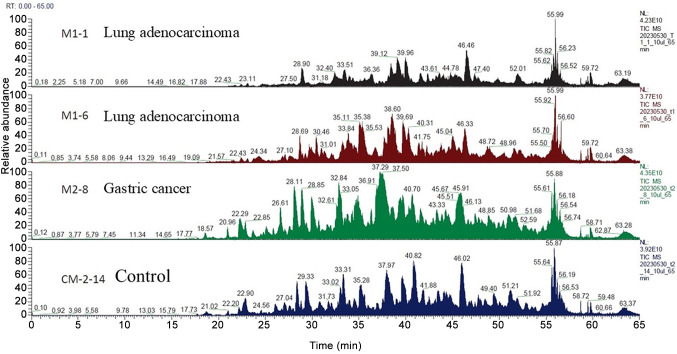
Patients and methods: A pilot study was performed using the multivariate analysis method and principal component analysis-discriminant analysis on proteomic results obtained using liquid chromatography-mass spectrometry (LC/MS) in two patients with lung cancer, one patient with gastric cancer, and one healthy control individual.
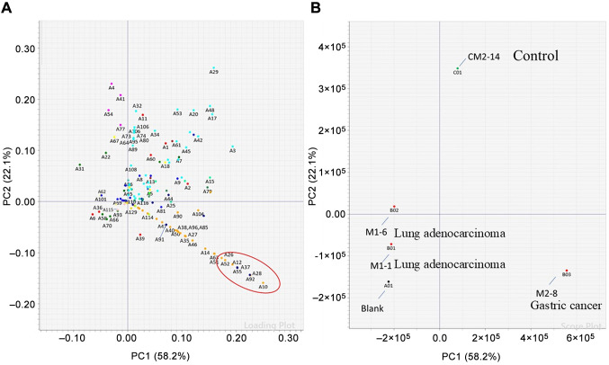
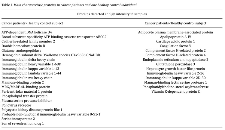
Results: No differences in specific proteins associated with high CEA levels were detected between lung and gastric cancers using LC/MS. Therefore, we performed statistical analysis using principal component analysis-discriminant analysis to determine whether there were differences in the protein signal patterns obtained using LC/MS. The results showed that the plots obtained for each patient and the healthy control were located in different quadrants of the four-quadrant matrix scatter plot.
Conclusion: Our results suggest the possibility of visually differentiating the primary tumor site in patients with elevated CEA levels. This method may also help recognize false-positive CEA results.
期刊介绍:
IN VIVO is an international peer-reviewed journal designed to bring together original high quality works and reviews on experimental and clinical biomedical research within the frames of physiology, pathology and disease management.
The topics of IN VIVO include: 1. Experimental development and application of new diagnostic and therapeutic procedures; 2. Pharmacological and toxicological evaluation of new drugs, drug combinations and drug delivery systems; 3. Clinical trials; 4. Development and characterization of models of biomedical research; 5. Cancer diagnosis and treatment; 6. Immunotherapy and vaccines; 7. Radiotherapy, Imaging; 8. Tissue engineering, Regenerative medicine; 9. Carcinogenesis.

 求助内容:
求助内容: 应助结果提醒方式:


