{"title":"Effect of Zoledronic Acid Administration Timing on Metastatic Bone Tumors.","authors":"Manabu Watanabe, Hiroyuki Tsuchie, Hiroyuki Nagasawa, Michio Hongo, Yuji Kasukawa, Daisuke Kudo, Fumihito Kasama, Keita Oya, Takashi Kawaragi, Naohisa Miyakoshi","doi":"10.21873/invivo.13995","DOIUrl":null,"url":null,"abstract":"<p><strong>Background/aim: </strong>Breast cancer frequently metastasizes to the bone, reducing quality of life and survival. Zoledronic acid (ZA), is used to treat bone metastasis; however, differences in efficacy depending on the timing of administration are not clear. This study investigated the effects of different timing of ZA administration in a mouse model of breast cancer bone metastasis.</p><p><strong>Materials and methods: </strong>E0771 cells (1.0×10<sup>5</sup> cells/10 μl) were injected into the femur of C57BL/6 mice to create a local bone metastasis model. The groups that started ZA administration one week before, at the same time as, one week after, and two weeks after tumor-cell administration were designated as the -1w, 0w, 1w, and 2w groups, respectively. A fifth group that did not receive ZA treatment was created as a control. ZA was administered at a dose of 100 μg/kg, and the same dose was administered once a week from the start of administration. The animals were sacrificed two and five weeks after tumor-cell administration. We evaluated body weight at the time of tumor-cell administration and sacrifice, and after sacrifice, the weight of the affected thigh, tumor volume, and bone destruction rate were determined using micro-computed tomography. Tumor necrosis and tumor growth were measured using histological immunostaining.</p><p><strong>Results: </strong>Five weeks after tumor-cell administration, bone destruction rate was significantly lower in all groups compared to the control group (<i>p</i><0.05). Additionally, the -1w group exhibited a significantly lower bone destruction rate than 1w and 2w groups (<i>p</i><0.05). There were no significant differences in tumor necrosis, but tumor growth was significantly lower in the -1w and 0w groups (<i>p</i><0.05).</p><p><strong>Conclusion: </strong>The earlier ZA was administered, the more strongly it suppressed bone destruction and tumor cell proliferation.</p>","PeriodicalId":13364,"journal":{"name":"In vivo","volume":"39 4","pages":"1984-1991"},"PeriodicalIF":1.8000,"publicationDate":"2025-07-01","publicationTypes":"Journal Article","fieldsOfStudy":null,"isOpenAccess":false,"openAccessPdf":"https://www.ncbi.nlm.nih.gov/pmc/articles/PMC12223653/pdf/","citationCount":"0","resultStr":null,"platform":"Semanticscholar","paperid":null,"PeriodicalName":"In vivo","FirstCategoryId":"3","ListUrlMain":"https://doi.org/10.21873/invivo.13995","RegionNum":4,"RegionCategory":"医学","ArticlePicture":[],"TitleCN":null,"AbstractTextCN":null,"PMCID":null,"EPubDate":"","PubModel":"","JCR":"Q3","JCRName":"MEDICINE, RESEARCH & EXPERIMENTAL","Score":null,"Total":0}
引用次数: 0
Abstract
Background/aim: Breast cancer frequently metastasizes to the bone, reducing quality of life and survival. Zoledronic acid (ZA), is used to treat bone metastasis; however, differences in efficacy depending on the timing of administration are not clear. This study investigated the effects of different timing of ZA administration in a mouse model of breast cancer bone metastasis.
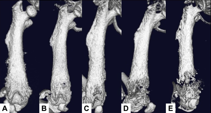
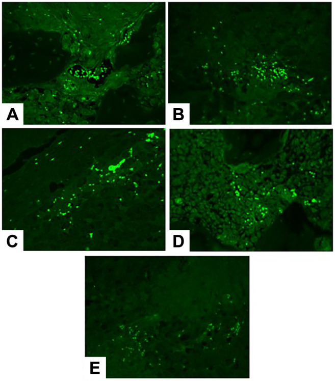
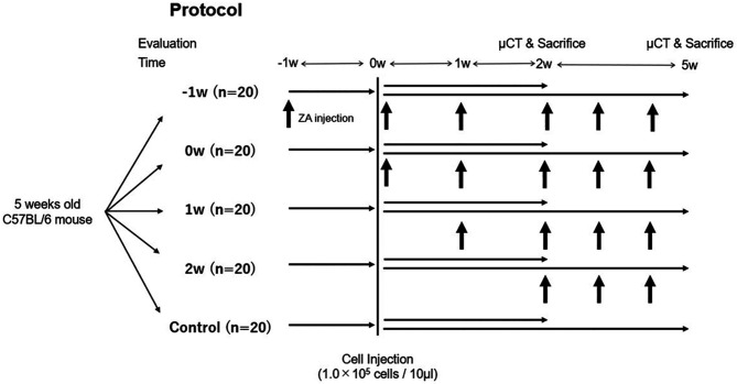
Materials and methods: E0771 cells (1.0×10⁵ cells/10 μl) were injected into the femur of C57BL/6 mice to create a local bone metastasis model. The groups that started ZA administration one week before, at the same time as, one week after, and two weeks after tumor-cell administration were designated as the -1w, 0w, 1w, and 2w groups, respectively. A fifth group that did not receive ZA treatment was created as a control. ZA was administered at a dose of 100 μg/kg, and the same dose was administered once a week from the start of administration. The animals were sacrificed two and five weeks after tumor-cell administration. We evaluated body weight at the time of tumor-cell administration and sacrifice, and after sacrifice, the weight of the affected thigh, tumor volume, and bone destruction rate were determined using micro-computed tomography. Tumor necrosis and tumor growth were measured using histological immunostaining.
Results: Five weeks after tumor-cell administration, bone destruction rate was significantly lower in all groups compared to the control group (p<0.05). Additionally, the -1w group exhibited a significantly lower bone destruction rate than 1w and 2w groups (p<0.05). There were no significant differences in tumor necrosis, but tumor growth was significantly lower in the -1w and 0w groups (p<0.05).
Conclusion: The earlier ZA was administered, the more strongly it suppressed bone destruction and tumor cell proliferation.
期刊介绍:
IN VIVO is an international peer-reviewed journal designed to bring together original high quality works and reviews on experimental and clinical biomedical research within the frames of physiology, pathology and disease management.
The topics of IN VIVO include: 1. Experimental development and application of new diagnostic and therapeutic procedures; 2. Pharmacological and toxicological evaluation of new drugs, drug combinations and drug delivery systems; 3. Clinical trials; 4. Development and characterization of models of biomedical research; 5. Cancer diagnosis and treatment; 6. Immunotherapy and vaccines; 7. Radiotherapy, Imaging; 8. Tissue engineering, Regenerative medicine; 9. Carcinogenesis.

 求助内容:
求助内容: 应助结果提醒方式:


