Surraj Susai, Mrudula Chandrupatla, Sumitra Sivakoti, Govindrao N Kusneniwa, Anand K Pyati
{"title":"Clumpy Novel Mitochondrial Signatures in Irradiated Human Diabetic Buccal Cells: A Case Control Study.","authors":"Surraj Susai, Mrudula Chandrupatla, Sumitra Sivakoti, Govindrao N Kusneniwa, Anand K Pyati","doi":"10.4274/MMJ.galenos.2025.87120","DOIUrl":null,"url":null,"abstract":"<p><strong>Objective: </strong>This study aimed to determine the whole free mitochondria in type-2 diabetic buccal epithelial cells using a supravital stain called \"Janus Green B\" and assess their behavior after exposure to near infrared light. The researchers observed microscopic mitochondrial load and its intracellular spatial behavior after exposure to near-infrared rays, bridging the gap in understanding mitochondrial orientation in diseases like diabetes. The aim of this research was to find out the quantitative involvement and electrostatic intracellular spatial patterns of whole mitochondria in diabetes.</p><p><strong>Methods: </strong>Exfoliated buccal cell wet mounts, supravitally stained using Janus green, and excited using infrared rays, were observed using an advanced bright field Axiocam microscope, and the images of whole mitochondria within the cells were photographed and analyzed using the ZEN 2.0 cell sense software. The migration patterns of mitochondria were observed.</p><p><strong>Results: </strong>A quantitative decrease in mitochondria was noted in diabetic cells. Signatures of clumpy peripheral shifts in mitochondria were observed in diabetic buccal cells post radiation.</p><p><strong>Conclusions: </strong>Advanced glycation end products of diabetes combined with oxidative stressors influenced the free mitochondria to clump peripherally and produce a characteristic signature. The decreased mitochondrial load contributed additional evidence to the reduced respiratory capacity of cells, which forced mitochondria to emit a detectable signature when irradiated.</p>","PeriodicalId":37427,"journal":{"name":"Medeniyet medical journal","volume":"40 2","pages":"93-100"},"PeriodicalIF":1.1000,"publicationDate":"2025-06-26","publicationTypes":"Journal Article","fieldsOfStudy":null,"isOpenAccess":false,"openAccessPdf":"https://www.ncbi.nlm.nih.gov/pmc/articles/PMC12203440/pdf/","citationCount":"0","resultStr":null,"platform":"Semanticscholar","paperid":null,"PeriodicalName":"Medeniyet medical journal","FirstCategoryId":"1085","ListUrlMain":"https://doi.org/10.4274/MMJ.galenos.2025.87120","RegionNum":0,"RegionCategory":null,"ArticlePicture":[],"TitleCN":null,"AbstractTextCN":null,"PMCID":null,"EPubDate":"","PubModel":"","JCR":"Q2","JCRName":"MEDICINE, GENERAL & INTERNAL","Score":null,"Total":0}
引用次数: 0
Abstract
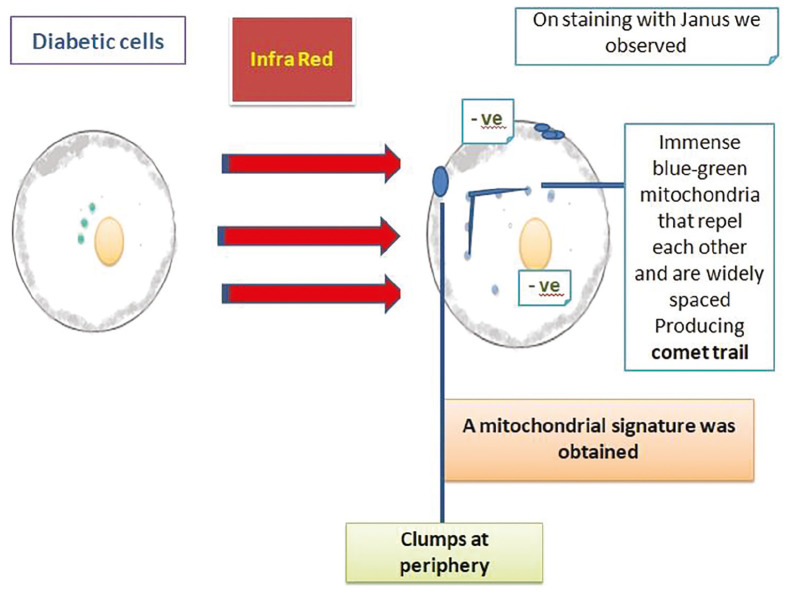
Objective: This study aimed to determine the whole free mitochondria in type-2 diabetic buccal epithelial cells using a supravital stain called "Janus Green B" and assess their behavior after exposure to near infrared light. The researchers observed microscopic mitochondrial load and its intracellular spatial behavior after exposure to near-infrared rays, bridging the gap in understanding mitochondrial orientation in diseases like diabetes. The aim of this research was to find out the quantitative involvement and electrostatic intracellular spatial patterns of whole mitochondria in diabetes.
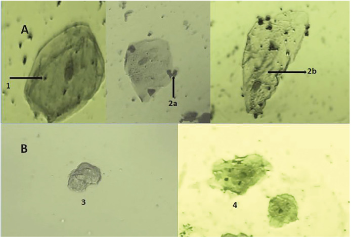
Methods: Exfoliated buccal cell wet mounts, supravitally stained using Janus green, and excited using infrared rays, were observed using an advanced bright field Axiocam microscope, and the images of whole mitochondria within the cells were photographed and analyzed using the ZEN 2.0 cell sense software. The migration patterns of mitochondria were observed.
Results: A quantitative decrease in mitochondria was noted in diabetic cells. Signatures of clumpy peripheral shifts in mitochondria were observed in diabetic buccal cells post radiation.
Conclusions: Advanced glycation end products of diabetes combined with oxidative stressors influenced the free mitochondria to clump peripherally and produce a characteristic signature. The decreased mitochondrial load contributed additional evidence to the reduced respiratory capacity of cells, which forced mitochondria to emit a detectable signature when irradiated.
目的:利用“Janus Green B”染色法检测2型糖尿病口腔上皮细胞的游离线粒体,并评估其在近红外光照射下的行为。研究人员观察了近红外线照射后的微观线粒体负荷及其细胞内空间行为,弥合了对糖尿病等疾病中线粒体取向的理解差距。本研究的目的是发现全线粒体在糖尿病中的定量参与和静电胞内空间格局。方法:取脱落的颊部细胞湿载片,表面采用Janus绿染色,红外线激发,采用先进的明场Axiocam显微镜观察,并用ZEN 2.0细胞感知软件对细胞内全线粒体图像进行拍摄和分析。观察线粒体的迁移模式。结果:糖尿病细胞线粒体数量减少。在糖尿病口腔细胞放射后观察到线粒体的团块外周移位的特征。结论:糖尿病晚期糖基化终产物结合氧化应激影响游离线粒体外周聚集并产生特征性特征。线粒体负荷的减少为细胞呼吸能力的降低提供了额外的证据,这迫使线粒体在辐射时发出可检测的信号。
期刊介绍:
The Medeniyet Medical Journal (Medeniyet Med J) is an open access, peer-reviewed, and scientific journal of Istanbul Medeniyet University Faculty of Medicine on various academic disciplines in medicine, which is published in English four times a year, in March, June, September, and December by a group of academics. Medeniyet Medical Journal is the continuation of Göztepe Medical Journal (ISSN: 1300-526X) which was started publishing in 1985. It changed the name as Medeniyet Medical Journal in 2015. Submission and publication are free of charge. No fees are asked from the authors for evaluation or publication process. All published articles are available online in the journal website (www.medeniyetmedicaljournal.org) without any fee. The journal publishes intradisciplinary or interdisciplinary clinical, experimental, and basic researches as well as original case reports, reviews, invited reviews, or letters to the editor, Being published since 1985, the Medeniyet Med J recognizes that the best science should lead to better lives based on the fact that the medicine should serve to the needs of society, and knowledge should transform society. The journal aims to address current issues at both national and international levels, start debates, and exert an influence on decision-makers all over the world by integrating science in everyday life. Medeniyet Med J is committed to serve the public and influence people’s lives in a positive way by making science widely accessible. Believing that the only goal is improving lives, and research has an impact on people’s lives, we select the best research papers in line with this goal.

 求助内容:
求助内容: 应助结果提醒方式:


