{"title":"EEA for sellar chodrosarcomas: case series with literature review.","authors":"GuoFo Ma, Ning Qiao, Wentao Wu, BoChao Zhang, Kefan Cai, SongBai Gui","doi":"10.1186/s41016-025-00397-4","DOIUrl":null,"url":null,"abstract":"<p><p>BACKGROUND : Chondrosarcoma is a rare and malignant tumor in the sellar region. Due to the limited understanding of its oncological behavior, it is often misdiagnosed as other lesions, such as chordoma or invasive pituitary adenoma. In the past, craniotomy was considered the primary treatment option. However, with the advancement of neuroendoscopic techniques, many centers have begun adopting endoscopic approaches for this disease. In this article, we summarized our case series and reviewed the previous papers to evaluate the clinical outcomes of neuroendoscopic resection for sellar chondrosarcomas.</p><p><strong>Methods: </strong>Four patients with sellar chondrosarcomas who underwent tumor resection by endonasal endoscopic approach (EEA) in our institute from 2017 to 2021 were reviewed. In addition, we reviewed the current literatures on sellar chondrosarcomas.</p><p><strong>Results: </strong>Our series included 4 patients, and 8 cases of sellar chondrosarcomas were reported in previous literatures. In the pooled cohort, there were 6 males and 6 females. The median age at diagnosis was 28.5 years (interquartile range, 22.8-39.3). The most common clinical presentation was blurring of vision (66.7%) and headache (50%). The incidence of preoperative endocrine disorder in such patients was 33.3%; these abnormalities could return to normal after surgery. Complete resection and incomplete resection were achieved in 7 (58.3%) and 5 (41.7%) cases, respectively. Among the 12 patients, only one patient was diagnosed with chondrosarcoma preoperatively; other patients were misdiagnosed with chordoma (n = 5; 41.7%), invasive non-functioning pituitary adenoma (INPA) (n = 4; 33.3%), or craniopharyngioma (n = 2; 16.7%).</p><p><strong>Conclusions: </strong>The preoperative diagnosis of sellar chondrosarcoma remains challenging and often requires differentiation from chordoma, INPA, or craniopharyngioma. When a calcified mass in the sellar region presents with intact or slightly disturbed anterior pituitary function, heterogeneous enhancement and no diffusion restriction on Magnetic Resonance Imaging(MRI) sequences, and surrounding bony destruction and bony attachment on Computed Tomography(CT) scans, a chondrosarcoma should be suspected preferentially. Complete resection is the optimal goal for the management of sellar chondrosarcoma, but adjuvant radiotherapy and periodic follow-up should be highlighted.</p>","PeriodicalId":36700,"journal":{"name":"Chinese Neurosurgical Journal","volume":"11 1","pages":"13"},"PeriodicalIF":0.0000,"publicationDate":"2025-06-26","publicationTypes":"Journal Article","fieldsOfStudy":null,"isOpenAccess":false,"openAccessPdf":"https://www.ncbi.nlm.nih.gov/pmc/articles/PMC12199512/pdf/","citationCount":"0","resultStr":null,"platform":"Semanticscholar","paperid":null,"PeriodicalName":"Chinese Neurosurgical Journal","FirstCategoryId":"3","ListUrlMain":"https://doi.org/10.1186/s41016-025-00397-4","RegionNum":0,"RegionCategory":null,"ArticlePicture":[],"TitleCN":null,"AbstractTextCN":null,"PMCID":null,"EPubDate":"","PubModel":"","JCR":"Q2","JCRName":"Medicine","Score":null,"Total":0}
引用次数: 0
Abstract
BACKGROUND : Chondrosarcoma is a rare and malignant tumor in the sellar region. Due to the limited understanding of its oncological behavior, it is often misdiagnosed as other lesions, such as chordoma or invasive pituitary adenoma. In the past, craniotomy was considered the primary treatment option. However, with the advancement of neuroendoscopic techniques, many centers have begun adopting endoscopic approaches for this disease. In this article, we summarized our case series and reviewed the previous papers to evaluate the clinical outcomes of neuroendoscopic resection for sellar chondrosarcomas.
Methods: Four patients with sellar chondrosarcomas who underwent tumor resection by endonasal endoscopic approach (EEA) in our institute from 2017 to 2021 were reviewed. In addition, we reviewed the current literatures on sellar chondrosarcomas.
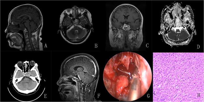
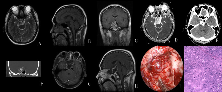
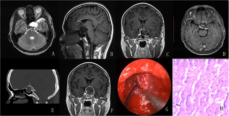
Results: Our series included 4 patients, and 8 cases of sellar chondrosarcomas were reported in previous literatures. In the pooled cohort, there were 6 males and 6 females. The median age at diagnosis was 28.5 years (interquartile range, 22.8-39.3). The most common clinical presentation was blurring of vision (66.7%) and headache (50%). The incidence of preoperative endocrine disorder in such patients was 33.3%; these abnormalities could return to normal after surgery. Complete resection and incomplete resection were achieved in 7 (58.3%) and 5 (41.7%) cases, respectively. Among the 12 patients, only one patient was diagnosed with chondrosarcoma preoperatively; other patients were misdiagnosed with chordoma (n = 5; 41.7%), invasive non-functioning pituitary adenoma (INPA) (n = 4; 33.3%), or craniopharyngioma (n = 2; 16.7%).
Conclusions: The preoperative diagnosis of sellar chondrosarcoma remains challenging and often requires differentiation from chordoma, INPA, or craniopharyngioma. When a calcified mass in the sellar region presents with intact or slightly disturbed anterior pituitary function, heterogeneous enhancement and no diffusion restriction on Magnetic Resonance Imaging(MRI) sequences, and surrounding bony destruction and bony attachment on Computed Tomography(CT) scans, a chondrosarcoma should be suspected preferentially. Complete resection is the optimal goal for the management of sellar chondrosarcoma, but adjuvant radiotherapy and periodic follow-up should be highlighted.
 求助内容:
求助内容: 应助结果提醒方式:


