{"title":"Coexisting Remnants of the Omphalomesenteric Duct and Urachus in an Infant.","authors":"Noboru Oyachi, Fuminori Numano","doi":"10.70352/scrj.cr.25-0003","DOIUrl":null,"url":null,"abstract":"<p><strong>Introduction: </strong>Congenital anomalies of the umbilicus, including remnants of the omphalomesenteric duct and urachus, result from the incomplete regression of fetal structures around the 10th week of gestation. The coexistence of these anomalies in a single patient is exceptionally uncommon. This report presents the case of a neonate with an umbilical nodule and periumbilical cyst, subsequently identified as coexisting remnants of the omphalomesenteric duct and urachus.</p><p><strong>Case presentation: </strong>This study reports the case of a 17-day-old female infant who presented with a small moist umbilical nodule and a persistent yellowish mucinous discharge. Initial treatment for umbilical granuloma failed to resolve the lesion. Imaging revealed a 2-cm cyst beneath the umbilicus and a cord-like structure connecting it to the bladder. Surgical exploration identified a 6-cm fibrous band extending from the cyst to the ileal wall, consistent with an omphalomesenteric duct remnant, and a 5-mm diameter urachal remnant connecting the cyst to the bladder. Histological analysis confirmed the presence of intestinal mucosa and transitional epithelium. The postoperative recovery of the patient was without complications.</p><p><strong>Conclusions: </strong>This case elucidates the diagnostic challenges posed by persistent umbilical lesions and highlights the importance of detailed imaging and surgical exploration for identifying rare congenital anomalies. Histopathological confirmation is essential for an accurate diagnosis. Further research is required to clarify the embryological basis and clinical implications of these anomalies.</p>","PeriodicalId":22096,"journal":{"name":"Surgical Case Reports","volume":"11 1","pages":""},"PeriodicalIF":0.7000,"publicationDate":"2025-01-01","publicationTypes":"Journal Article","fieldsOfStudy":null,"isOpenAccess":false,"openAccessPdf":"https://www.ncbi.nlm.nih.gov/pmc/articles/PMC12197850/pdf/","citationCount":"0","resultStr":null,"platform":"Semanticscholar","paperid":null,"PeriodicalName":"Surgical Case Reports","FirstCategoryId":"1085","ListUrlMain":"https://doi.org/10.70352/scrj.cr.25-0003","RegionNum":0,"RegionCategory":null,"ArticlePicture":[],"TitleCN":null,"AbstractTextCN":null,"PMCID":null,"EPubDate":"2025/6/21 0:00:00","PubModel":"Epub","JCR":"Q4","JCRName":"SURGERY","Score":null,"Total":0}
引用次数: 0
Abstract
Introduction: Congenital anomalies of the umbilicus, including remnants of the omphalomesenteric duct and urachus, result from the incomplete regression of fetal structures around the 10th week of gestation. The coexistence of these anomalies in a single patient is exceptionally uncommon. This report presents the case of a neonate with an umbilical nodule and periumbilical cyst, subsequently identified as coexisting remnants of the omphalomesenteric duct and urachus.
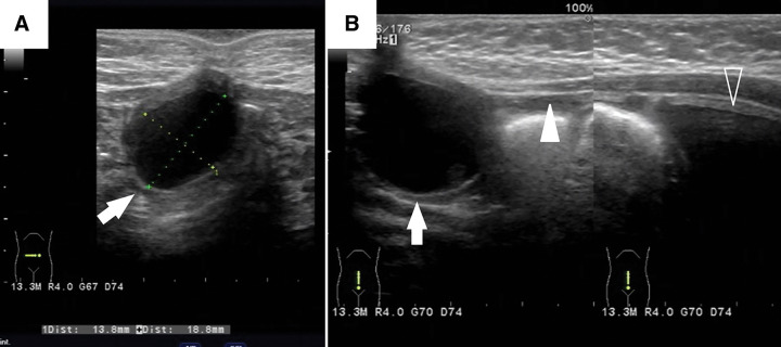
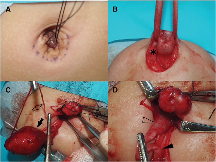
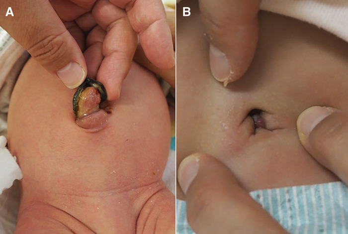
Case presentation: This study reports the case of a 17-day-old female infant who presented with a small moist umbilical nodule and a persistent yellowish mucinous discharge. Initial treatment for umbilical granuloma failed to resolve the lesion. Imaging revealed a 2-cm cyst beneath the umbilicus and a cord-like structure connecting it to the bladder. Surgical exploration identified a 6-cm fibrous band extending from the cyst to the ileal wall, consistent with an omphalomesenteric duct remnant, and a 5-mm diameter urachal remnant connecting the cyst to the bladder. Histological analysis confirmed the presence of intestinal mucosa and transitional epithelium. The postoperative recovery of the patient was without complications.
Conclusions: This case elucidates the diagnostic challenges posed by persistent umbilical lesions and highlights the importance of detailed imaging and surgical exploration for identifying rare congenital anomalies. Histopathological confirmation is essential for an accurate diagnosis. Further research is required to clarify the embryological basis and clinical implications of these anomalies.

 求助内容:
求助内容: 应助结果提醒方式:


