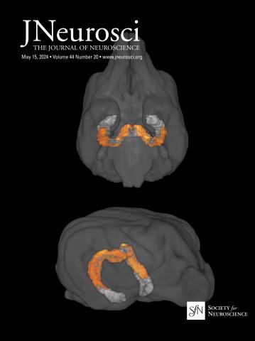
Pre-microsaccadic modulation in foveal V1: enhancement in the current and future stimulus locations.
IF 4.4
2区 医学
Q1 NEUROSCIENCES
引用次数: 0
Abstract
Microsaccades are miniature saccades performed during visual fixation that were shown to play a pivotal role in active sensing. Recent studies suggested that pre-microsaccadic attention may underlie the enhanced visual processing at the stimulus site. However, the neuronal mechanism underlying this phenomenon at the foveal scale remains unknown. Using voltage-sensitive dye imaging we investigated the neural responses to uninstructed, spontaneous microsaccades in the fovea of the primary visual cortex (V1) in behaving monkeys (macaque, male). We found that prior to microsaccades onset toward a small visual stimulus, the neuronal activity at the current and future landing stimulus sites was enhanced relative to microsaccades away from the stimulus. This enhancement was spatially confined to the current and future landing stimulus sites, which appeared to merge along the microsaccades ( < 1 deg ) trajectory in V1. Finally, we found a pre-microsaccadic increased synchronization at the current stimulus site. Our findings shed new light on neural modulations preceding microsaccades and suggest a link to neural signatures of attention.Significance statement Microsaccades are miniature eye-movements that occur during visual fixation. Behavioral studies have suggested that pre-microsaccadic attention enhances visual processing in the fovea. However, the underlying neuronal mechanisms at the foveal scale remain unknown. Using voltage-sensitive dye imaging in monkeys, we investigated how microsaccades influence neural activity in the foveal region of the primary visual cortex. Just before a microsaccade toward a small visual stimulus, neural activity was enhanced at both the current and future landing stimulus locations, compared to microsaccades directed away. This enhancement appeared over the microsaccade path and was accompanied by increased synchronization at the current stimulus location. Our findings reveal novel neural modulations preceding microsaccades, and suggest a link between microsaccades and neural signatures of attention.中央凹V1微囊前调制:当前和未来刺激位置的增强。
微眼跳是在视觉固定期间进行的微型眼跳,在主动感知中起着关键作用。最近的研究表明,微眼球前注意可能是刺激部位视觉加工增强的基础。然而,这种现象背后的神经元机制在中央凹尺度上仍然未知。使用电压敏感染料成像技术,我们研究了行为猴子(雄性猕猴)初级视觉皮层中央凹(V1)对无指示、自发微跳的神经反应。我们发现,在朝向小视觉刺激的微扫视开始之前,当前和未来着陆刺激点的神经元活动相对于远离刺激的微扫视增强。这种增强在空间上仅限于当前和未来的着陆刺激位点,它们似乎沿着V1的微跳(< 1度)轨迹合并。最后,我们发现在当前的刺激部位,微跳前同步增加。我们的发现为微跳前的神经调节提供了新的线索,并提出了与注意力的神经特征的联系。微扫视是在注视过程中发生的微小眼球运动。行为研究表明,微眼球前注意增强了中央凹的视觉处理。然而,在中央凹尺度上的潜在神经元机制仍然未知。在猴子中使用电压敏感染料成像,我们研究了微眼跳如何影响初级视觉皮层中央凹区域的神经活动。在微扫视朝向一个小的视觉刺激之前,与微扫视朝向其他地方相比,当前和未来着陆刺激点的神经活动都增强了。这种增强出现在微跃动路径上,并伴随着当前刺激位置同步性的增强。我们的研究结果揭示了微跳前新的神经调节,并提出了微跳和注意力的神经特征之间的联系。
本文章由计算机程序翻译,如有差异,请以英文原文为准。
求助全文
约1分钟内获得全文
求助全文
来源期刊

Journal of Neuroscience
医学-神经科学
CiteScore
9.30
自引率
3.80%
发文量
1164
审稿时长
12 months
期刊介绍:
JNeurosci (ISSN 0270-6474) is an official journal of the Society for Neuroscience. It is published weekly by the Society, fifty weeks a year, one volume a year. JNeurosci publishes papers on a broad range of topics of general interest to those working on the nervous system. Authors now have an Open Choice option for their published articles
 求助内容:
求助内容: 应助结果提醒方式:


