Utility of a Modified Penlight-Cover Test for Neurolocalization of Lesions Based on Visual Suppression of Nystagmus in Dogs and Cats With Vestibular Disease
Alice Chan, Gemma E. Longson, Edward Ives, Claire Turner, Paul Freeman, Stacey Brady, Ana Martinez Loro, Bruno Scalia, Susana Monforte Monteiro, Sara Formoso, Sam Khan, An E. Vanhaesebrouck
{"title":"Utility of a Modified Penlight-Cover Test for Neurolocalization of Lesions Based on Visual Suppression of Nystagmus in Dogs and Cats With Vestibular Disease","authors":"Alice Chan, Gemma E. Longson, Edward Ives, Claire Turner, Paul Freeman, Stacey Brady, Ana Martinez Loro, Bruno Scalia, Susana Monforte Monteiro, Sara Formoso, Sam Khan, An E. Vanhaesebrouck","doi":"10.1111/jvim.70182","DOIUrl":null,"url":null,"abstract":"<div>\n \n \n <section>\n \n <h3> Background</h3>\n \n <p>Humans with peripheral vestibular disorders can suppress nystagmus through visual fixation, a capability often compromised in those with central vestibular disorders. Bedside tests that exploit this difference can aid neurolocalization in humans. These tests remain unexplored in veterinary medicine.</p>\n </section>\n \n <section>\n \n <h3> Hypothesis</h3>\n \n <p>Removal of visual input will reveal or enhance nystagmus in animals with peripheral vestibular disease, while animals with central vestibular disease would show little change.</p>\n </section>\n \n <section>\n \n <h3> Animals</h3>\n \n <p>Twenty-one dogs and cats with peripheral vestibular lesions and 16 with central vestibular lesions. Diagnosis was confirmed by MRI.</p>\n </section>\n \n <section>\n \n <h3> Methods</h3>\n \n <p>A prospective study was conducted using a modified penlight-cover test. Because animals cannot be easily instructed to fixate on a visual target, removal of visual input was used as a substitute for eliminating visual fixation, based on the assumption that visual fixation also occurs spontaneously. A 0.5-W LED penlight was shined into one eye while covering the other to eliminate visual input. Nystagmus beat frequency (BF) and subjective evaluation of slow phase velocity (SPV) were recorded before and during penlight application.</p>\n </section>\n \n <section>\n \n <h3> Results</h3>\n \n <p>In animals with peripheral lesions, BF increased in 33% and SPV in 24% of cases after removal of visual input. Among those with central lesions, only one of 16 showed an increase in BF, and none exhibited an increase in SPV.</p>\n </section>\n \n <section>\n \n <h3> Conclusions</h3>\n \n <p>When used alongside the neurological examination, the modified penlight-cover test, could raise suspicion of a peripheral vestibular lesion if it reveals increased BF or SPV.</p>\n </section>\n </div>","PeriodicalId":49958,"journal":{"name":"Journal of Veterinary Internal Medicine","volume":"39 4","pages":""},"PeriodicalIF":2.2000,"publicationDate":"2025-06-27","publicationTypes":"Journal Article","fieldsOfStudy":null,"isOpenAccess":false,"openAccessPdf":"https://onlinelibrary.wiley.com/doi/epdf/10.1111/jvim.70182","citationCount":"0","resultStr":null,"platform":"Semanticscholar","paperid":null,"PeriodicalName":"Journal of Veterinary Internal Medicine","FirstCategoryId":"97","ListUrlMain":"https://onlinelibrary.wiley.com/doi/10.1111/jvim.70182","RegionNum":2,"RegionCategory":"农林科学","ArticlePicture":[],"TitleCN":null,"AbstractTextCN":null,"PMCID":null,"EPubDate":"","PubModel":"","JCR":"Q1","JCRName":"VETERINARY SCIENCES","Score":null,"Total":0}
引用次数: 0
Abstract
Background
Humans with peripheral vestibular disorders can suppress nystagmus through visual fixation, a capability often compromised in those with central vestibular disorders. Bedside tests that exploit this difference can aid neurolocalization in humans. These tests remain unexplored in veterinary medicine.
Hypothesis
Removal of visual input will reveal or enhance nystagmus in animals with peripheral vestibular disease, while animals with central vestibular disease would show little change.
Animals
Twenty-one dogs and cats with peripheral vestibular lesions and 16 with central vestibular lesions. Diagnosis was confirmed by MRI.
Methods
A prospective study was conducted using a modified penlight-cover test. Because animals cannot be easily instructed to fixate on a visual target, removal of visual input was used as a substitute for eliminating visual fixation, based on the assumption that visual fixation also occurs spontaneously. A 0.5-W LED penlight was shined into one eye while covering the other to eliminate visual input. Nystagmus beat frequency (BF) and subjective evaluation of slow phase velocity (SPV) were recorded before and during penlight application.
Results
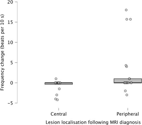
In animals with peripheral lesions, BF increased in 33% and SPV in 24% of cases after removal of visual input. Among those with central lesions, only one of 16 showed an increase in BF, and none exhibited an increase in SPV.
Conclusions
When used alongside the neurological examination, the modified penlight-cover test, could raise suspicion of a peripheral vestibular lesion if it reveals increased BF or SPV.
期刊介绍:
The mission of the Journal of Veterinary Internal Medicine is to advance veterinary medical knowledge and improve the lives of animals by publication of authoritative scientific articles of animal diseases.
 求助内容:
求助内容: 应助结果提醒方式:


