Juan Ángel Aibar-Duran, Rodrigo Akira Watanabe, Nathan J McDannold, G Rees Cosgrove
{"title":"New technique for direct targeting of the ventral intermediate nucleus using magnetic resonance-guided focused ultrasound.","authors":"Juan Ángel Aibar-Duran, Rodrigo Akira Watanabe, Nathan J McDannold, G Rees Cosgrove","doi":"10.3389/fradi.2025.1588379","DOIUrl":null,"url":null,"abstract":"<p><strong>Background: </strong>Accurate targeting and lesion placement are critical in treating movement disorders with magnetic resonance-guided focused ultrasound (MRgFUS). Indirect atlas-based targeting often lacks precision. Direct anatomical targeting with 3T MRI offers a promising alternative. This report aims to refine MRgFUS thalamotomy by integrating advanced imaging and lesion conformality strategies.</p><p><strong>Material and methods: </strong>Preoperative and postoperative white matter null (WMn) MR-imaging was employed for direct Vim detection. Essential anatomical landmarks are identified. Dual-lesion conformality was used to adapt to the spatial distribution of the Vim.</p><p><strong>Results: </strong>Lesions of the Vim were identifiable using the postoperative WMn MRI sequence. The direct visualization of the Vim usually avoids extension into the internal capsule and the sensory thalamic nucleus. Sagittal imaging confirmed the dual-lesion conformational strategy which conforms to the antero-superior orientation of the Vim.</p><p><strong>Conclusions: </strong>Direct Vim targeting for MRgFUS is feasible for individual cases with the use of WMnMPRAGE MRI sequences. The use of lesion conformality adapts well to the anatomical and spatial distribution of Vim. Further studies will be needed to confirm the safety profile of this approach and correlate with clinical outcomes.</p>","PeriodicalId":73101,"journal":{"name":"Frontiers in radiology","volume":"5 ","pages":"1588379"},"PeriodicalIF":2.3000,"publicationDate":"2025-06-11","publicationTypes":"Journal Article","fieldsOfStudy":null,"isOpenAccess":false,"openAccessPdf":"https://www.ncbi.nlm.nih.gov/pmc/articles/PMC12187783/pdf/","citationCount":"0","resultStr":null,"platform":"Semanticscholar","paperid":null,"PeriodicalName":"Frontiers in radiology","FirstCategoryId":"1085","ListUrlMain":"https://doi.org/10.3389/fradi.2025.1588379","RegionNum":0,"RegionCategory":null,"ArticlePicture":[],"TitleCN":null,"AbstractTextCN":null,"PMCID":null,"EPubDate":"2025/1/1 0:00:00","PubModel":"eCollection","JCR":"","JCRName":"","Score":null,"Total":0}
引用次数: 0
Abstract
Background: Accurate targeting and lesion placement are critical in treating movement disorders with magnetic resonance-guided focused ultrasound (MRgFUS). Indirect atlas-based targeting often lacks precision. Direct anatomical targeting with 3T MRI offers a promising alternative. This report aims to refine MRgFUS thalamotomy by integrating advanced imaging and lesion conformality strategies.
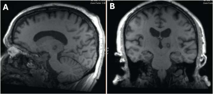
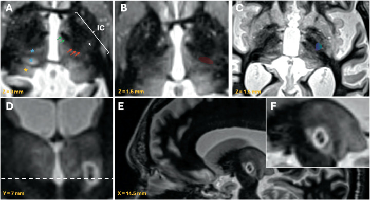
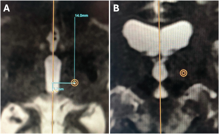
Material and methods: Preoperative and postoperative white matter null (WMn) MR-imaging was employed for direct Vim detection. Essential anatomical landmarks are identified. Dual-lesion conformality was used to adapt to the spatial distribution of the Vim.
Results: Lesions of the Vim were identifiable using the postoperative WMn MRI sequence. The direct visualization of the Vim usually avoids extension into the internal capsule and the sensory thalamic nucleus. Sagittal imaging confirmed the dual-lesion conformational strategy which conforms to the antero-superior orientation of the Vim.
Conclusions: Direct Vim targeting for MRgFUS is feasible for individual cases with the use of WMnMPRAGE MRI sequences. The use of lesion conformality adapts well to the anatomical and spatial distribution of Vim. Further studies will be needed to confirm the safety profile of this approach and correlate with clinical outcomes.

 求助内容:
求助内容: 应助结果提醒方式:


