Massimo Cesareo, Alessio Martucci, Roberta Bovenzi, Marco Lombardo, Francesca Pistoia, Vittoria Carla D'Agostino, Alessandro Stefani, Carlo Nucci, Nicola Biagio Mercuri, Maria Albanese
{"title":"Evaluating the impact of anti-CGRP monoclonal antibodies on retinal features in migraine patients: a retrospective optical coherence tomography study.","authors":"Massimo Cesareo, Alessio Martucci, Roberta Bovenzi, Marco Lombardo, Francesca Pistoia, Vittoria Carla D'Agostino, Alessandro Stefani, Carlo Nucci, Nicola Biagio Mercuri, Maria Albanese","doi":"10.1177/17562864251347277","DOIUrl":null,"url":null,"abstract":"<p><strong>Background: </strong>Migraine is a disabling neurovascular disorder characterized by recurrent attacks that lead to extracranial and visual involvement. Several studies have investigated the retinal vasculature features in individuals with migraine, but there have been conflicting results.</p><p><strong>Objective: </strong>To evaluate retinal structure in migraine patients before (T0) and after 6-month therapy (T1) with anti-calcitonin gene-related peptide (CGRP) monoclonal antibodies (mAbs), using optical coherence tomography (OCT) imaging.</p><p><strong>Design: </strong>A case-control and longitudinal study was conducted between January 2021 and December 2023, including 20 eyes from 10 healthy controls (HCs) and 32 eyes of 16 patients with migraine and treated with anti-CGRP mAbs according to AIFA criteria.</p><p><strong>Methods: </strong>Patients underwent OCT angiography (OCT-A) to assess retinal vessel density (VD) and spectral-domain OCT (SD-OCT) to evaluate central retinal thickness, macular structure, and peripapillary retinal nerve fiber layer thickness. These parameters were assessed in both groups at T0 and again after 6 months (T1) as part of routine clinical care.</p><p><strong>Results: </strong>All migraineurs exhibited a significant reduction in disease disability at T1, as assessed by clinical parameters. OCT data analysis revealed that individuals with migraine showed a significant increase in temporal retinal nerve fiber layer (RNFL) thickness and a reduction in nasal RNFL thickness compared to HCs. No differences in retinal circulation were observed between the groups at baseline. At T1, RNFL thickness remained sustained in the superior temporal sector, while the percentage VD of the superficial capillary plexus and radial peripapillary capillary significantly increased in the nasal perifoveal, inferior temporal, and hemi-inferior subregions.</p><p><strong>Conclusion: </strong>Our study suggests that specific retinal structural changes could precede vascular dysfunction in migraine and can be detected early by combining SD-OCT and OCT-A acquisitions. Short-term treatment with anti-CGRP mAbs may exert neuroprotective effects, potentially preventing permanent ocular damage.</p><p><strong>Trial registration: </strong>EyeHEAD Study (Trial registration number AIFA July/2024: IT 1735, www.aifa.gov.it/registro-studi-osservazionali).</p>","PeriodicalId":22980,"journal":{"name":"Therapeutic Advances in Neurological Disorders","volume":"18 ","pages":"17562864251347277"},"PeriodicalIF":4.1000,"publicationDate":"2025-06-24","publicationTypes":"Journal Article","fieldsOfStudy":null,"isOpenAccess":false,"openAccessPdf":"https://www.ncbi.nlm.nih.gov/pmc/articles/PMC12188072/pdf/","citationCount":"0","resultStr":null,"platform":"Semanticscholar","paperid":null,"PeriodicalName":"Therapeutic Advances in Neurological Disorders","FirstCategoryId":"3","ListUrlMain":"https://doi.org/10.1177/17562864251347277","RegionNum":2,"RegionCategory":"医学","ArticlePicture":[],"TitleCN":null,"AbstractTextCN":null,"PMCID":null,"EPubDate":"2025/1/1 0:00:00","PubModel":"eCollection","JCR":"Q1","JCRName":"CLINICAL NEUROLOGY","Score":null,"Total":0}
引用次数: 0
Abstract
Background: Migraine is a disabling neurovascular disorder characterized by recurrent attacks that lead to extracranial and visual involvement. Several studies have investigated the retinal vasculature features in individuals with migraine, but there have been conflicting results.
Objective: To evaluate retinal structure in migraine patients before (T0) and after 6-month therapy (T1) with anti-calcitonin gene-related peptide (CGRP) monoclonal antibodies (mAbs), using optical coherence tomography (OCT) imaging.
Design: A case-control and longitudinal study was conducted between January 2021 and December 2023, including 20 eyes from 10 healthy controls (HCs) and 32 eyes of 16 patients with migraine and treated with anti-CGRP mAbs according to AIFA criteria.
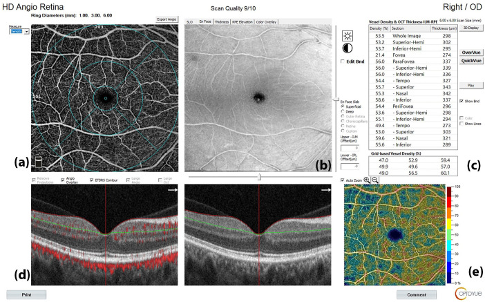
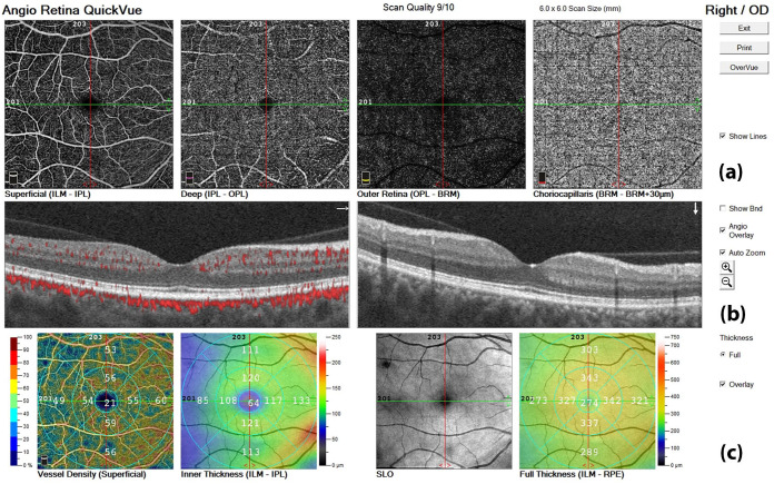
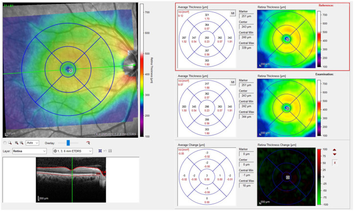
Methods: Patients underwent OCT angiography (OCT-A) to assess retinal vessel density (VD) and spectral-domain OCT (SD-OCT) to evaluate central retinal thickness, macular structure, and peripapillary retinal nerve fiber layer thickness. These parameters were assessed in both groups at T0 and again after 6 months (T1) as part of routine clinical care.
Results: All migraineurs exhibited a significant reduction in disease disability at T1, as assessed by clinical parameters. OCT data analysis revealed that individuals with migraine showed a significant increase in temporal retinal nerve fiber layer (RNFL) thickness and a reduction in nasal RNFL thickness compared to HCs. No differences in retinal circulation were observed between the groups at baseline. At T1, RNFL thickness remained sustained in the superior temporal sector, while the percentage VD of the superficial capillary plexus and radial peripapillary capillary significantly increased in the nasal perifoveal, inferior temporal, and hemi-inferior subregions.
Conclusion: Our study suggests that specific retinal structural changes could precede vascular dysfunction in migraine and can be detected early by combining SD-OCT and OCT-A acquisitions. Short-term treatment with anti-CGRP mAbs may exert neuroprotective effects, potentially preventing permanent ocular damage.
Trial registration: EyeHEAD Study (Trial registration number AIFA July/2024: IT 1735, www.aifa.gov.it/registro-studi-osservazionali).
期刊介绍:
Therapeutic Advances in Neurological Disorders is a peer-reviewed, open access journal delivering the highest quality articles, reviews, and scholarly comment on pioneering efforts and innovative studies across all areas of neurology. The journal has a strong clinical and pharmacological focus and is aimed at clinicians and researchers in neurology, providing a forum in print and online for publishing the highest quality articles in this area.

 求助内容:
求助内容: 应助结果提醒方式:


