A Rare Case of Cervical Solitary Fibrous Tumor in a Pediatric Patient: Case Report and Literature Review.
IF 2
Q3 CLINICAL NEUROLOGY
引用次数: 0
Abstract
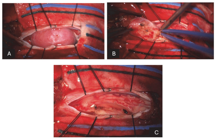
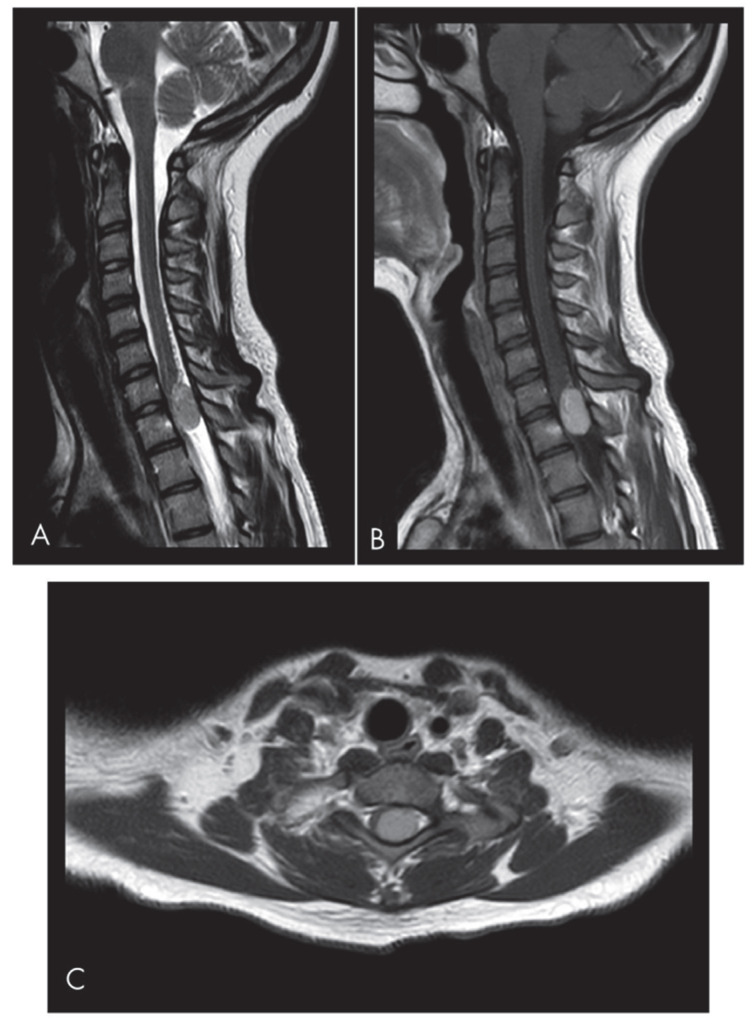
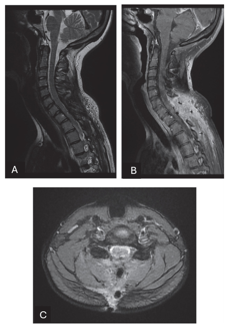
Solitary fibrous tumors (SFTs) are rare mesenchymal neoplasms of fibroblastic origin. In this study, we report a rare case of cervical SFT in a pediatric patient, a rare phenomenon since the incidence is particularly rare in pediatric patients according to the literature. Typical radiological features of the lesion may lead to misdiagnosis. Image study and immunohistochemistry are crucial for its correct diagnosis. Their imaging characteristics often resemble meningiomas or schwannomas, making differential diagnosis challenging. Immunohistochemical markers such as CD34 and STAT6 remain essential for definitive diagnosis.
小儿宫颈孤立性纤维性肿瘤1例:病例报告及文献复习。
孤立性纤维性肿瘤是一种罕见的纤维母细胞间充质肿瘤。在本研究中,我们报告了一例罕见的小儿颈椎SFT病例,这是一种罕见的现象,因为根据文献,小儿患者的发病率尤其罕见。典型的放射学特征可能导致误诊。影像检查和免疫组化对其正确诊断至关重要。其影像学特征常与脑膜瘤或神经鞘瘤相似,难以鉴别诊断。免疫组织化学标志物如CD34和STAT6仍然是明确诊断的必要条件。
本文章由计算机程序翻译,如有差异,请以英文原文为准。
求助全文
约1分钟内获得全文
求助全文

 求助内容:
求助内容: 应助结果提醒方式:


