{"title":"Presumed Solitary Circumscribed Retinal Astrocytic Proliferation: A Case Report.","authors":"Mojtaba Abrishami, Daniel J Weisbrod, Hatem Krema","doi":"10.4103/joco.joco_136_24","DOIUrl":null,"url":null,"abstract":"<p><strong>Purpose: </strong>To report a case of presumed solitary circumscribed retinal astrocytic proliferation (PSCRAP) diagnosed through multimodal imaging.</p><p><strong>Methods: </strong>A single case report documented with multimodal imaging.</p><p><strong>Results: </strong>A 41-year-old asymptomatic female was referred for a second opinion regarding whitish retinal lesions in her left eye, first identified by her local optometrist 8 years prior. Her history included Hashimoto thyroiditis, with no personal or family history of tuberous sclerosis complex or neurofibromatosis. Visual acuity was 20/20 in both eyes. Fundoscopy of the left eye revealed two pearly white avascular retinal masses. Spectral-domain optical coherence tomography (SD-OCT) showed hyperreflective intraretinal masses with optical shadowing. Optical coherence tomography angiography (OCTA) demonstrated a signal void in the retinal vascular plexuses. Fundus autofluorescence showed moderate hyperautofluorescence. The lesions remained stable over 3 months.</p><p><strong>Conclusions: </strong>PSCRAP is a rare, benign retinal tumor. Multimodal imaging, including SD-OCT and OCTA, is essential for accurate diagnosis, showing unique features such as separation from the retinal nerve fiber layer and lack of intrinsic vascularity. Our observation of two lesions raises questions about the solitary nature of this condition. Continued documentation may be necessary to differentiate from simulating lesions that may undergo subsequent growth.</p>","PeriodicalId":15423,"journal":{"name":"Journal of Current Ophthalmology","volume":"36 3","pages":"296-299"},"PeriodicalIF":0.9000,"publicationDate":"2025-06-05","publicationTypes":"Journal Article","fieldsOfStudy":null,"isOpenAccess":false,"openAccessPdf":"https://www.ncbi.nlm.nih.gov/pmc/articles/PMC12184867/pdf/","citationCount":"0","resultStr":null,"platform":"Semanticscholar","paperid":null,"PeriodicalName":"Journal of Current Ophthalmology","FirstCategoryId":"1085","ListUrlMain":"https://doi.org/10.4103/joco.joco_136_24","RegionNum":0,"RegionCategory":null,"ArticlePicture":[],"TitleCN":null,"AbstractTextCN":null,"PMCID":null,"EPubDate":"2024/7/1 0:00:00","PubModel":"eCollection","JCR":"Q3","JCRName":"OPHTHALMOLOGY","Score":null,"Total":0}
引用次数: 0
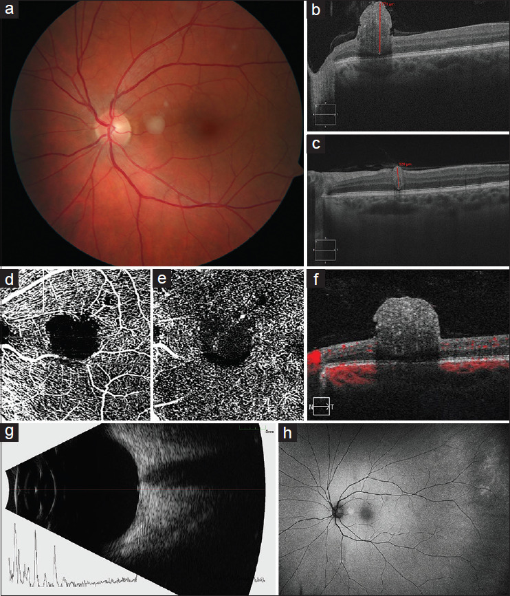
Abstract
Purpose: To report a case of presumed solitary circumscribed retinal astrocytic proliferation (PSCRAP) diagnosed through multimodal imaging.
Methods: A single case report documented with multimodal imaging.
Results: A 41-year-old asymptomatic female was referred for a second opinion regarding whitish retinal lesions in her left eye, first identified by her local optometrist 8 years prior. Her history included Hashimoto thyroiditis, with no personal or family history of tuberous sclerosis complex or neurofibromatosis. Visual acuity was 20/20 in both eyes. Fundoscopy of the left eye revealed two pearly white avascular retinal masses. Spectral-domain optical coherence tomography (SD-OCT) showed hyperreflective intraretinal masses with optical shadowing. Optical coherence tomography angiography (OCTA) demonstrated a signal void in the retinal vascular plexuses. Fundus autofluorescence showed moderate hyperautofluorescence. The lesions remained stable over 3 months.
Conclusions: PSCRAP is a rare, benign retinal tumor. Multimodal imaging, including SD-OCT and OCTA, is essential for accurate diagnosis, showing unique features such as separation from the retinal nerve fiber layer and lack of intrinsic vascularity. Our observation of two lesions raises questions about the solitary nature of this condition. Continued documentation may be necessary to differentiate from simulating lesions that may undergo subsequent growth.
期刊介绍:
Peer Review under the responsibility of Iranian Society of Ophthalmology Journal of Current Ophthalmology, the official publication of the Iranian Society of Ophthalmology, is a peer-reviewed, open-access, scientific journal that welcomes high quality original articles related to vision science and all fields of ophthalmology. Journal of Current Ophthalmology is the continuum of Iranian Journal of Ophthalmology published since 1969.

 求助内容:
求助内容: 应助结果提醒方式:


