Hemichorea Associated With Nigrostriatal Dysfunction: Case Report of a Patient With an Ipsilateral Infarct in the Lenticular Nucleus and Internal Capsule.
{"title":"Hemichorea Associated With Nigrostriatal Dysfunction: Case Report of a Patient With an Ipsilateral Infarct in the Lenticular Nucleus and Internal Capsule.","authors":"Makoto Kobayashi","doi":"10.1155/crnm/6054686","DOIUrl":null,"url":null,"abstract":"<p><p>Hemichorea is a rare manifestation of ischemic stroke whose lesion is typically located in the contralateral basal ganglia. Its pathomechanism has not been elucidated completely; however, it may be related to nigrostriatal dysfunction. In patients with hemichorea, dopamine transporter-single photon emission computed tomography (DAT-SPECT) reportedly displayed decreased tracer accumulation in the contralateral striatum. Moreover, in exceptional cases, responsible lesions were located in the ipsilateral cerebral hemisphere. This case report describes an 84-year-old man who presented with three weeks of intermittent, involuntary, and twisting movements in his right limbs. On physical examination, the patient had right-sided hemichorea without other neurological deficits. The choreic movements were more frequent in the lower limb than in the upper and provoked when he tried to take a certain posture or engaged in mental arithmetic. Magnetic resonance imaging performed on suspicion of stroke detected a right hemispheric subacute infarct in the posterior part of the lenticular nucleus and posterior limb of the internal capsule. Furthermore, DAT-SPECT revealed decreased tracer accumulation in the right striatum. He was administered oral antiplatelet medication after being diagnosed with lacunar infarction. The choreic movements gradually reduced over the next 8 months and eventually disappeared. The lesion in the lenticular nucleus and internal capsule was considered to have induced ipsilesional hemichorea, considering the temporal proximity between the hemichorea and ischemic stroke. Although DAT-SPECT findings in patients with ipsilesional hemichorea have not been reported, this case suggests that nigrostriatal dopamine dysfunction can contribute to the pathogenesis of ipsilesional hemichorea.</p>","PeriodicalId":9615,"journal":{"name":"Case Reports in Neurological Medicine","volume":"2025 ","pages":"6054686"},"PeriodicalIF":0.9000,"publicationDate":"2025-06-17","publicationTypes":"Journal Article","fieldsOfStudy":null,"isOpenAccess":false,"openAccessPdf":"https://www.ncbi.nlm.nih.gov/pmc/articles/PMC12187432/pdf/","citationCount":"0","resultStr":null,"platform":"Semanticscholar","paperid":null,"PeriodicalName":"Case Reports in Neurological Medicine","FirstCategoryId":"1085","ListUrlMain":"https://doi.org/10.1155/crnm/6054686","RegionNum":0,"RegionCategory":null,"ArticlePicture":[],"TitleCN":null,"AbstractTextCN":null,"PMCID":null,"EPubDate":"2025/1/1 0:00:00","PubModel":"eCollection","JCR":"Q4","JCRName":"CLINICAL NEUROLOGY","Score":null,"Total":0}
引用次数: 0
Abstract
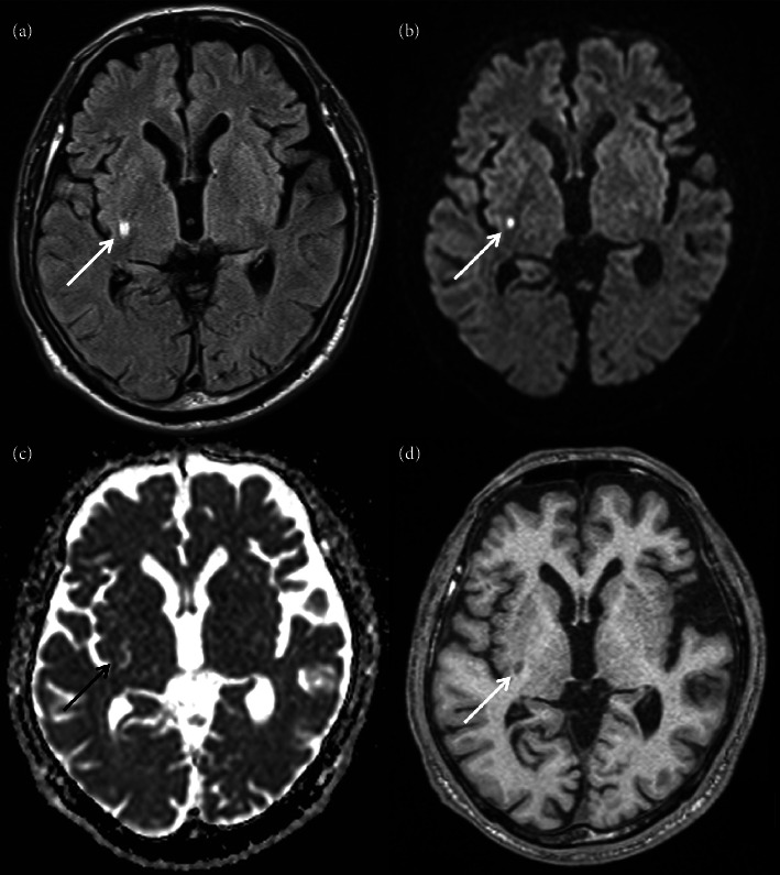
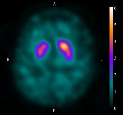
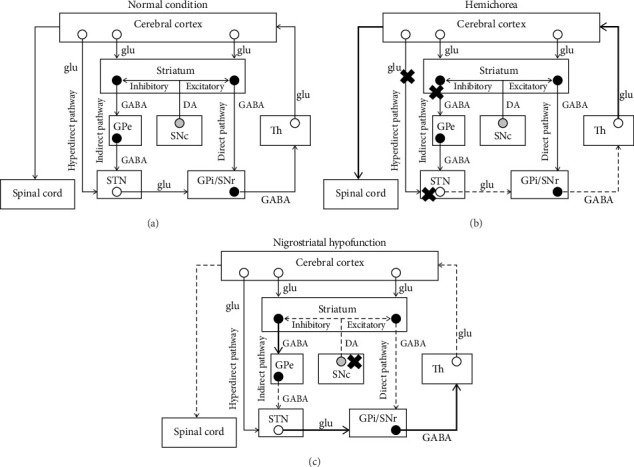
Hemichorea is a rare manifestation of ischemic stroke whose lesion is typically located in the contralateral basal ganglia. Its pathomechanism has not been elucidated completely; however, it may be related to nigrostriatal dysfunction. In patients with hemichorea, dopamine transporter-single photon emission computed tomography (DAT-SPECT) reportedly displayed decreased tracer accumulation in the contralateral striatum. Moreover, in exceptional cases, responsible lesions were located in the ipsilateral cerebral hemisphere. This case report describes an 84-year-old man who presented with three weeks of intermittent, involuntary, and twisting movements in his right limbs. On physical examination, the patient had right-sided hemichorea without other neurological deficits. The choreic movements were more frequent in the lower limb than in the upper and provoked when he tried to take a certain posture or engaged in mental arithmetic. Magnetic resonance imaging performed on suspicion of stroke detected a right hemispheric subacute infarct in the posterior part of the lenticular nucleus and posterior limb of the internal capsule. Furthermore, DAT-SPECT revealed decreased tracer accumulation in the right striatum. He was administered oral antiplatelet medication after being diagnosed with lacunar infarction. The choreic movements gradually reduced over the next 8 months and eventually disappeared. The lesion in the lenticular nucleus and internal capsule was considered to have induced ipsilesional hemichorea, considering the temporal proximity between the hemichorea and ischemic stroke. Although DAT-SPECT findings in patients with ipsilesional hemichorea have not been reported, this case suggests that nigrostriatal dopamine dysfunction can contribute to the pathogenesis of ipsilesional hemichorea.

 求助内容:
求助内容: 应助结果提醒方式:


