Mohamed A. Abdel Hamid, Samir A. Elborolosy, Sara El Moshy, Hany R. Ammar, S. Sivasankaran, Walid S. Salem, Elham A. Hassan
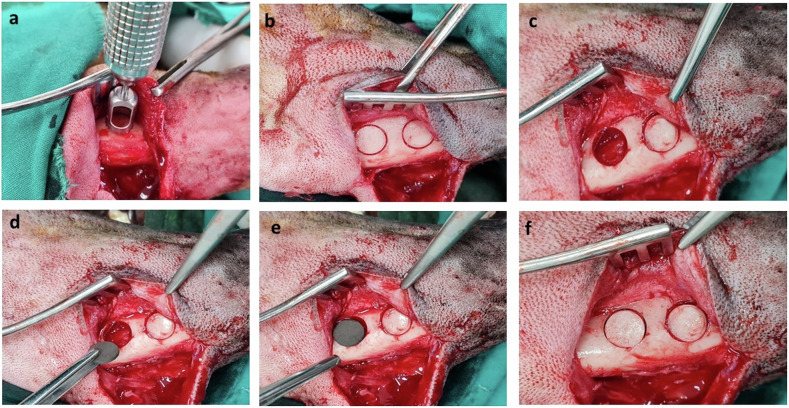
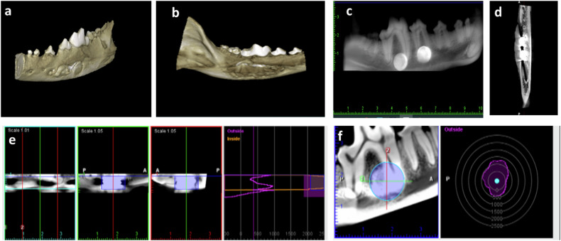
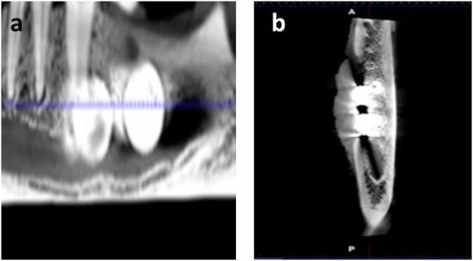
{"title":"Osteogenic potential of nanostructured Cu/W/Co in Fe-Mn alloys designed for maxillofacial applications: in vivo study in dog model","authors":"Mohamed A. Abdel Hamid, Samir A. Elborolosy, Sara El Moshy, Hany R. Ammar, S. Sivasankaran, Walid S. Salem, Elham A. Hassan","doi":"10.1007/s10856-025-06890-7","DOIUrl":null,"url":null,"abstract":"<div><p>To investigate osteogenic potential of biodegradable nanostructured Cu/W/Co in Fe-Mn alloys for maxillofacial applications in an in vivo model. Nanostructured FeMn<sub>35</sub>, FeMn<sub>32</sub>Cu<sub>3</sub>, FeMn<sub>32</sub>W<sub>3,</sub> FeMn<sub>32</sub>Co<sub>3</sub> alloys were fabricated. Ten mongrel dogs were included where five mandibular defects were induced in each dog. Defects were randomly allocated into 5 groups ((M) control defects covered by bone disc, (M0) implanted by FeMn<sub>35</sub> alloy, (M1) implanted by FeMn<sub>32</sub>Cu<sub>3</sub> alloy, (M2) implanted by FeMn<sub>32</sub>W<sub>3</sub> alloy, (M3) implanted by FeMn<sub>32</sub>Co<sub>3</sub> alloy). Dogs were euthanized at 12 weeks for cone beam computed tomography, histologic and immunohistologic evaluation, and gene expression of osteopontin and osteocalcin. Defects implanted with metal demonstrated thicker bone trabeculae mixed with lamellar bone while control defects (M) demonestrated immature woven bone. Quantitative evaluation of bone area %, area % of mature bone and expression of osteopentin and osteocalcin bone markers revealed a statistically significant highest bone area % and maturation in group M3 compared to M2, M1, M0, and M group. A statistically significant increase in bone area % and maturation was recorded in M2 group compared to M1, M0, and M groups. A significantly increased bone area % and maturation was recorded in M1 compared to control M group and also between M0 and M group. Incorporating Cu/W/Co into Fe-Mn alloys enhanced biocompatibility and improved bone regeneration suggesting its suitability for use in various orthopedic and dental applications. Biodegradable metal alloys could improve patient outcome, reduce the need for additional surgeries to remove nonbiodegradble implants. Biodegradable metal alloys could improve patient outcome, reduce the need for additional surgeries to remove nonbiodegradble implants.</p><h3>Graphical Abstract</h3><div><figure><div><div><picture><source><img></source></picture></div></div></figure></div></div>","PeriodicalId":647,"journal":{"name":"Journal of Materials Science: Materials in Medicine","volume":"36 1","pages":""},"PeriodicalIF":4.5000,"publicationDate":"2025-06-25","publicationTypes":"Journal Article","fieldsOfStudy":null,"isOpenAccess":false,"openAccessPdf":"https://www.ncbi.nlm.nih.gov/pmc/articles/PMC12198315/pdf/","citationCount":"0","resultStr":null,"platform":"Semanticscholar","paperid":null,"PeriodicalName":"Journal of Materials Science: Materials in Medicine","FirstCategoryId":"5","ListUrlMain":"https://link.springer.com/article/10.1007/s10856-025-06890-7","RegionNum":3,"RegionCategory":"医学","ArticlePicture":[],"TitleCN":null,"AbstractTextCN":null,"PMCID":null,"EPubDate":"","PubModel":"","JCR":"Q2","JCRName":"ENGINEERING, BIOMEDICAL","Score":null,"Total":0}
引用次数: 0
Abstract
To investigate osteogenic potential of biodegradable nanostructured Cu/W/Co in Fe-Mn alloys for maxillofacial applications in an in vivo model. Nanostructured FeMn35, FeMn32Cu3, FeMn32W3, FeMn32Co3 alloys were fabricated. Ten mongrel dogs were included where five mandibular defects were induced in each dog. Defects were randomly allocated into 5 groups ((M) control defects covered by bone disc, (M0) implanted by FeMn35 alloy, (M1) implanted by FeMn32Cu3 alloy, (M2) implanted by FeMn32W3 alloy, (M3) implanted by FeMn32Co3 alloy). Dogs were euthanized at 12 weeks for cone beam computed tomography, histologic and immunohistologic evaluation, and gene expression of osteopontin and osteocalcin. Defects implanted with metal demonstrated thicker bone trabeculae mixed with lamellar bone while control defects (M) demonestrated immature woven bone. Quantitative evaluation of bone area %, area % of mature bone and expression of osteopentin and osteocalcin bone markers revealed a statistically significant highest bone area % and maturation in group M3 compared to M2, M1, M0, and M group. A statistically significant increase in bone area % and maturation was recorded in M2 group compared to M1, M0, and M groups. A significantly increased bone area % and maturation was recorded in M1 compared to control M group and also between M0 and M group. Incorporating Cu/W/Co into Fe-Mn alloys enhanced biocompatibility and improved bone regeneration suggesting its suitability for use in various orthopedic and dental applications. Biodegradable metal alloys could improve patient outcome, reduce the need for additional surgeries to remove nonbiodegradble implants. Biodegradable metal alloys could improve patient outcome, reduce the need for additional surgeries to remove nonbiodegradble implants.
期刊介绍:
The Journal of Materials Science: Materials in Medicine publishes refereed papers providing significant progress in the application of biomaterials and tissue engineering constructs as medical or dental implants, prostheses and devices. Coverage spans a wide range of topics from basic science to clinical applications, around the theme of materials in medicine and dentistry. The central element is the development of synthetic and natural materials used in orthopaedic, maxillofacial, cardiovascular, neurological, ophthalmic and dental applications. Special biomedical topics include biomaterial synthesis and characterisation, biocompatibility studies, nanomedicine, tissue engineering constructs and cell substrates, regenerative medicine, computer modelling and other advanced experimental methodologies.
 求助内容:
求助内容: 应助结果提醒方式:


