George Triantafyllou, Panagiotis Papadopoulos-Manolarakis, George Tsakotos, Anastasia Triantafyllou, Maria Piagkou
{"title":"Persistent trigeminal artery variant functioning as a duplicate superior cerebellar artery.","authors":"George Triantafyllou, Panagiotis Papadopoulos-Manolarakis, George Tsakotos, Anastasia Triantafyllou, Maria Piagkou","doi":"10.1007/s00276-025-03673-1","DOIUrl":null,"url":null,"abstract":"<p><p>The cerebral arterial circle displays significant variation, with infrequent configurations often ascertained through contemporary imaging techniques. We present a sporadic case of a duplicated superior cerebellar artery (SCA) discovered during magnetic resonance angiography (MRA) of a 58-year-old female patient. Typical SCA emanates from the distal basilar artery (BA), whereas a duplicate one originates from the pre-cavernous segment of the internal carotid artery (ICA). This accessory vessel traveled parallel to the primary SCA, supplying a portion of its standard vascular territory. This distinctive configuration corresponds to a PTA variant featuring a duplicate SCA, a combination very rarely reported in existing literature. The variant above underscores the embryological origins of the cerebral arterial circle, particularly involving carotid-vertebrobasilar anastomoses. The existence of both a typical and a duplicate SCA constitutes an unprecedented configuration within literature. These vascular anomalies possess clinical significance, especially to neurovascular compression syndromes, such as trigeminal neuralgia, or during surgical and endovascular interventions.</p>","PeriodicalId":49461,"journal":{"name":"Surgical and Radiologic Anatomy","volume":"47 1","pages":"164"},"PeriodicalIF":1.2000,"publicationDate":"2025-06-23","publicationTypes":"Journal Article","fieldsOfStudy":null,"isOpenAccess":false,"openAccessPdf":"https://www.ncbi.nlm.nih.gov/pmc/articles/PMC12185625/pdf/","citationCount":"0","resultStr":null,"platform":"Semanticscholar","paperid":null,"PeriodicalName":"Surgical and Radiologic Anatomy","FirstCategoryId":"3","ListUrlMain":"https://doi.org/10.1007/s00276-025-03673-1","RegionNum":4,"RegionCategory":"医学","ArticlePicture":[],"TitleCN":null,"AbstractTextCN":null,"PMCID":null,"EPubDate":"","PubModel":"","JCR":"Q2","JCRName":"Medicine","Score":null,"Total":0}
引用次数: 0
Abstract
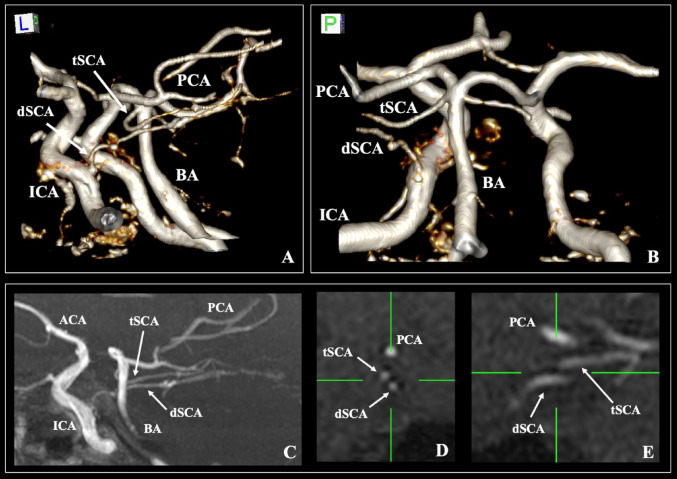
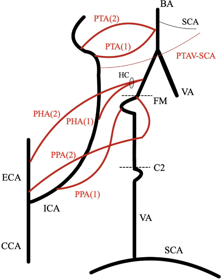
The cerebral arterial circle displays significant variation, with infrequent configurations often ascertained through contemporary imaging techniques. We present a sporadic case of a duplicated superior cerebellar artery (SCA) discovered during magnetic resonance angiography (MRA) of a 58-year-old female patient. Typical SCA emanates from the distal basilar artery (BA), whereas a duplicate one originates from the pre-cavernous segment of the internal carotid artery (ICA). This accessory vessel traveled parallel to the primary SCA, supplying a portion of its standard vascular territory. This distinctive configuration corresponds to a PTA variant featuring a duplicate SCA, a combination very rarely reported in existing literature. The variant above underscores the embryological origins of the cerebral arterial circle, particularly involving carotid-vertebrobasilar anastomoses. The existence of both a typical and a duplicate SCA constitutes an unprecedented configuration within literature. These vascular anomalies possess clinical significance, especially to neurovascular compression syndromes, such as trigeminal neuralgia, or during surgical and endovascular interventions.
期刊介绍:
Anatomy is a morphological science which cannot fail to interest the clinician. The practical application of anatomical research to clinical problems necessitates special adaptation and selectivity in choosing from numerous international works. Although there is a tendency to believe that meaningful advances in anatomy are unlikely, constant revision is necessary. Surgical and Radiologic Anatomy, the first international journal of Clinical anatomy has been created in this spirit.
Its goal is to serve clinicians, regardless of speciality-physicians, surgeons, radiologists or other specialists-as an indispensable aid with which they can improve their knowledge of anatomy. Each issue includes: Original papers, review articles, articles on the anatomical bases of medical, surgical and radiological techniques, articles of normal radiologic anatomy, brief reviews of anatomical publications of clinical interest.
Particular attention is given to high quality illustrations, which are indispensable for a better understanding of anatomical problems.
Surgical and Radiologic Anatomy is a journal written by anatomists for clinicians with a special interest in anatomy.

 求助内容:
求助内容: 应助结果提醒方式:


