{"title":"Complete remission of peritoneal strumosis from struma ovarii after radioiodine therapy: a case report.","authors":"Jun Yang, Zhengzhou Pan, Jili Wang, Xinhui Su","doi":"10.1186/s13044-025-00247-6","DOIUrl":null,"url":null,"abstract":"<p><strong>Background: </strong>Struma ovarii (SO) is a specialized monodermal teratoma composed predominantly of thyroid tissue (≥ 50%) and accounts for approximately 5% of all ovarian teratomas. In rare cases, the benign SO may spread to the peritoneal cavity and exhibit the histological features of struma ovarii in a condition termed peritoneal strumosis. Here, we present a rare case of complete remission of peritoneal strumosis from SO after radioiodine therapy.</p><p><strong>Case presentation: </strong>A 41-year-old Chinese woman underwent transabdominal left oophorectomy for a benign SO 18 years prior to presentation in the clinic. She was admitted to our institution for periodic medical examination after ultrasonography revealed a left pelvic mass. The patient underwent total abdominal hysterectomy, right salpingo-oophorectomy, and omentectomy, and multiple biopsies were taken from the omentum and mesentery. Pathology revealed peritoneal strumosis without evidence of malignancy from SO. Afterward, a total thyroidectomy was performed, and a histological examination revealed multinodular goiter. In total, 4400 MBq of 131I was administered, and lesion remission was confirmed. Finally, after 1 year of follow-up, the patient had no evidence of recurrence.</p><p><strong>Conclusion: </strong>Peritoneal strumosis from OS is a rare aggressive clinical manifestation that differs from malignancy. Conservative surgery with personalized radioiodine may be a practical therapeutic option for unresectable peritoneal strumosis, and long-term monitoring is recommended.</p>","PeriodicalId":39048,"journal":{"name":"Thyroid Research","volume":"18 1","pages":"29"},"PeriodicalIF":1.8000,"publicationDate":"2025-06-24","publicationTypes":"Journal Article","fieldsOfStudy":null,"isOpenAccess":false,"openAccessPdf":"https://www.ncbi.nlm.nih.gov/pmc/articles/PMC12186374/pdf/","citationCount":"0","resultStr":null,"platform":"Semanticscholar","paperid":null,"PeriodicalName":"Thyroid Research","FirstCategoryId":"1085","ListUrlMain":"https://doi.org/10.1186/s13044-025-00247-6","RegionNum":0,"RegionCategory":null,"ArticlePicture":[],"TitleCN":null,"AbstractTextCN":null,"PMCID":null,"EPubDate":"","PubModel":"","JCR":"Q3","JCRName":"ENDOCRINOLOGY & METABOLISM","Score":null,"Total":0}
引用次数: 0
Abstract
Background: Struma ovarii (SO) is a specialized monodermal teratoma composed predominantly of thyroid tissue (≥ 50%) and accounts for approximately 5% of all ovarian teratomas. In rare cases, the benign SO may spread to the peritoneal cavity and exhibit the histological features of struma ovarii in a condition termed peritoneal strumosis. Here, we present a rare case of complete remission of peritoneal strumosis from SO after radioiodine therapy.
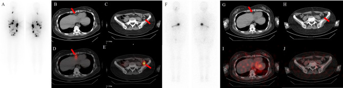
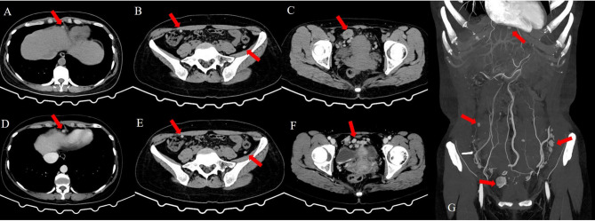
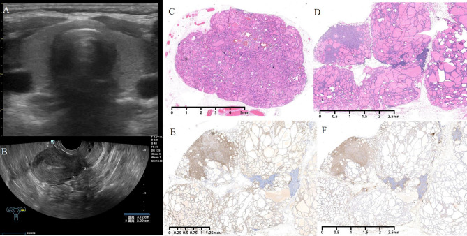
Case presentation: A 41-year-old Chinese woman underwent transabdominal left oophorectomy for a benign SO 18 years prior to presentation in the clinic. She was admitted to our institution for periodic medical examination after ultrasonography revealed a left pelvic mass. The patient underwent total abdominal hysterectomy, right salpingo-oophorectomy, and omentectomy, and multiple biopsies were taken from the omentum and mesentery. Pathology revealed peritoneal strumosis without evidence of malignancy from SO. Afterward, a total thyroidectomy was performed, and a histological examination revealed multinodular goiter. In total, 4400 MBq of 131I was administered, and lesion remission was confirmed. Finally, after 1 year of follow-up, the patient had no evidence of recurrence.
Conclusion: Peritoneal strumosis from OS is a rare aggressive clinical manifestation that differs from malignancy. Conservative surgery with personalized radioiodine may be a practical therapeutic option for unresectable peritoneal strumosis, and long-term monitoring is recommended.
 求助内容:
求助内容: 应助结果提醒方式:


