Benjamin Lüling, Fabian Preisner, Jeremias Motte, Anna Lena Fisse, Thomas Grüter, Rafael Klimas, Emelie Schäfer, Annika Altenborg, Devrim Colak, Jörg Philipps, Tim Godel, Daniel Schwarz, Sabine Heiland, Min-Suk Yoon, Ralf Gold, Martin Bendszus, Moritz Kronlage, Kalliopi Pitarokoili
{"title":"Comparison of imaging markers of nerve ultrasound and MR-neurography in a longitudinal course in chronic inflammatory demyelinating polyneuropathy.","authors":"Benjamin Lüling, Fabian Preisner, Jeremias Motte, Anna Lena Fisse, Thomas Grüter, Rafael Klimas, Emelie Schäfer, Annika Altenborg, Devrim Colak, Jörg Philipps, Tim Godel, Daniel Schwarz, Sabine Heiland, Min-Suk Yoon, Ralf Gold, Martin Bendszus, Moritz Kronlage, Kalliopi Pitarokoili","doi":"10.1177/17562864251342336","DOIUrl":null,"url":null,"abstract":"<p><strong>Background: </strong>The novel criteria for the diagnosis of chronic inflammatory demyelinating polyneuropathy (CIDP) have established imaging with nerve ultrasound (NUS) and magnetic resonance neurography (MRN) as complementary methods for CIDP diagnosis.</p><p><strong>Objectives: </strong>Our goal was to investigate the role of MRN and NUS for CIDP monitoring.</p><p><strong>Methods and design: </strong>We longitudinally examined 12 CIDP patients from 2016 to 2022 using NUS, MRN, nerve conduction studies (NCS), and clinical parameters (inflammatory neuropathy cause and treatment (INCAT)/overall disability sum score (ODSS)). NUS evaluated the cross-sectional area (CSA) of the median, ulnar, radial, tibial, fibular, and sural nerve as well as the intranerve CSA variability (INV<sub>csa</sub>) of the tibial, fibular, ulnar, and median nerve, whereas MRN evaluated T2-weighted sequences of the fibular and tibial nerve at the popliteal fossa.</p><p><strong>Results: </strong>Five patients showed clinical improvement/stability with corresponding improved or stable NCS/NUS parameters (number of nerves with increased CSA and INV<sub>CSA</sub>). Seven deteriorating patients showed deteriorating NCS and either increasing or decreasing NUS markers possibly indicating inflammatory activity or degenerative CSA reduction. The difference ΔINCAT/ODSS<sub>2022-2016</sub> correlated positively with NUS ΔINV<sub>CSA2022-2016</sub> (<i>p</i> = 0.007, <i>r</i> = 0.731, <i>n</i> = 12) and with NUS ΔCSA<sub>2022-2016</sub> of the tibial nerve (<i>p</i> = 0.0005, <i>r</i> = 0.865, <i>n</i> = 12). Further, NUS-CSA of the tibial nerve in the popliteal fossa in 2016 correlated inversely with the difference of the INCAT/ODSS score (ΔINCAT/ODSS<sub>2022-2016</sub>; <i>r</i> = -0.653; <i>p</i> = 0.033; <i>n</i> = 11). Finally, the Bland-Altman analyses for the tibial and fibular nerve showed a bias of -1.903 and 2.195 mm<sup>2</sup> (bias = NUS-CSA - MRN-CSA) accordingly revealing a difference between MRN and NUS measurements for deeper nerves.</p><p><strong>Conclusion: </strong>CSA and INV<sub>CSA</sub> of the tibial and fibular nerve can be used for monitoring in CIDP, and increased CSA of the tibial nerve is a good prognostic marker. MRN is more reliable for evaluating inflammation in proximal leg nerve segments.</p>","PeriodicalId":22980,"journal":{"name":"Therapeutic Advances in Neurological Disorders","volume":"18 ","pages":"17562864251342336"},"PeriodicalIF":4.1000,"publicationDate":"2025-06-21","publicationTypes":"Journal Article","fieldsOfStudy":null,"isOpenAccess":false,"openAccessPdf":"https://www.ncbi.nlm.nih.gov/pmc/articles/PMC12182616/pdf/","citationCount":"0","resultStr":null,"platform":"Semanticscholar","paperid":null,"PeriodicalName":"Therapeutic Advances in Neurological Disorders","FirstCategoryId":"3","ListUrlMain":"https://doi.org/10.1177/17562864251342336","RegionNum":2,"RegionCategory":"医学","ArticlePicture":[],"TitleCN":null,"AbstractTextCN":null,"PMCID":null,"EPubDate":"2025/1/1 0:00:00","PubModel":"eCollection","JCR":"Q1","JCRName":"CLINICAL NEUROLOGY","Score":null,"Total":0}
引用次数: 0
Abstract
Background: The novel criteria for the diagnosis of chronic inflammatory demyelinating polyneuropathy (CIDP) have established imaging with nerve ultrasound (NUS) and magnetic resonance neurography (MRN) as complementary methods for CIDP diagnosis.
Objectives: Our goal was to investigate the role of MRN and NUS for CIDP monitoring.
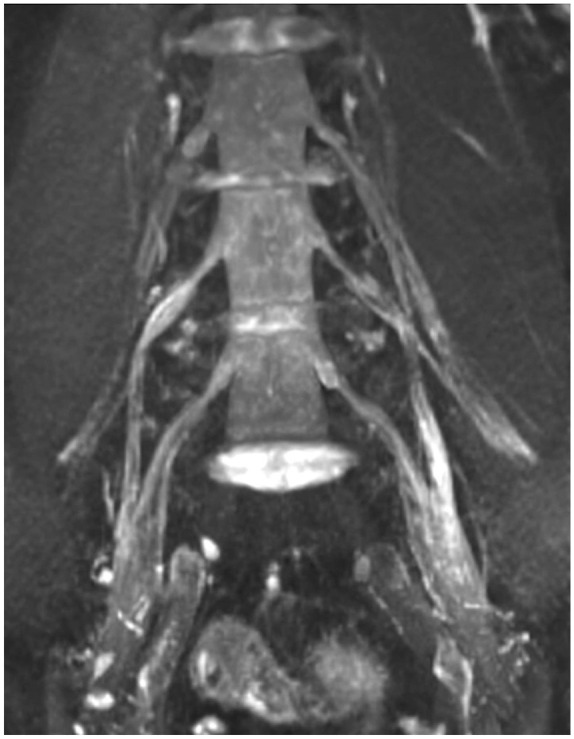
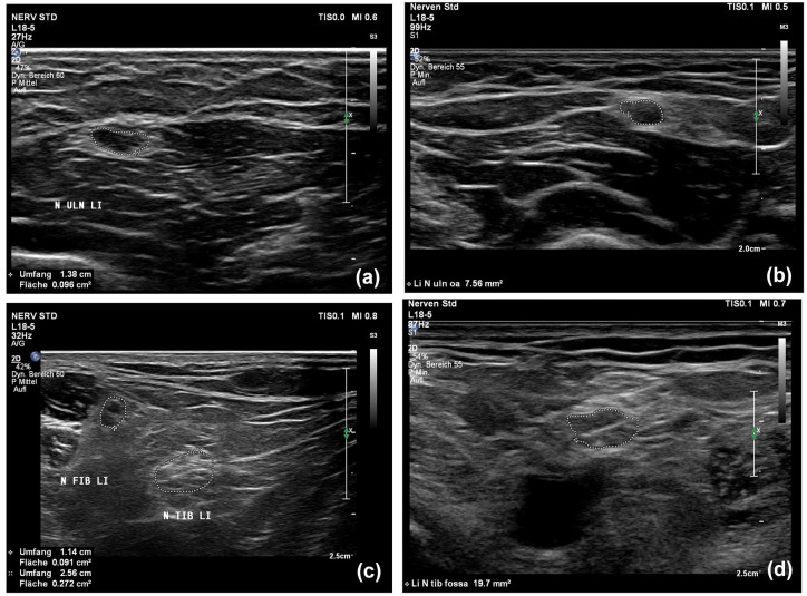
Methods and design: We longitudinally examined 12 CIDP patients from 2016 to 2022 using NUS, MRN, nerve conduction studies (NCS), and clinical parameters (inflammatory neuropathy cause and treatment (INCAT)/overall disability sum score (ODSS)). NUS evaluated the cross-sectional area (CSA) of the median, ulnar, radial, tibial, fibular, and sural nerve as well as the intranerve CSA variability (INVcsa) of the tibial, fibular, ulnar, and median nerve, whereas MRN evaluated T2-weighted sequences of the fibular and tibial nerve at the popliteal fossa.
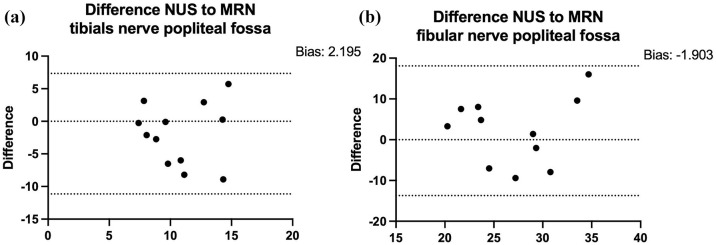
Results: Five patients showed clinical improvement/stability with corresponding improved or stable NCS/NUS parameters (number of nerves with increased CSA and INVCSA). Seven deteriorating patients showed deteriorating NCS and either increasing or decreasing NUS markers possibly indicating inflammatory activity or degenerative CSA reduction. The difference ΔINCAT/ODSS2022-2016 correlated positively with NUS ΔINVCSA2022-2016 (p = 0.007, r = 0.731, n = 12) and with NUS ΔCSA2022-2016 of the tibial nerve (p = 0.0005, r = 0.865, n = 12). Further, NUS-CSA of the tibial nerve in the popliteal fossa in 2016 correlated inversely with the difference of the INCAT/ODSS score (ΔINCAT/ODSS2022-2016; r = -0.653; p = 0.033; n = 11). Finally, the Bland-Altman analyses for the tibial and fibular nerve showed a bias of -1.903 and 2.195 mm2 (bias = NUS-CSA - MRN-CSA) accordingly revealing a difference between MRN and NUS measurements for deeper nerves.
Conclusion: CSA and INVCSA of the tibial and fibular nerve can be used for monitoring in CIDP, and increased CSA of the tibial nerve is a good prognostic marker. MRN is more reliable for evaluating inflammation in proximal leg nerve segments.
期刊介绍:
Therapeutic Advances in Neurological Disorders is a peer-reviewed, open access journal delivering the highest quality articles, reviews, and scholarly comment on pioneering efforts and innovative studies across all areas of neurology. The journal has a strong clinical and pharmacological focus and is aimed at clinicians and researchers in neurology, providing a forum in print and online for publishing the highest quality articles in this area.

 求助内容:
求助内容: 应助结果提醒方式:


