Omid Mirmosayyeb, Mohammad Yazdan Panah, Reza Kord, Elahe Espoo, Saeed Vaheb, Aram Zabeti, Vahid Shaygannejad
{"title":"Optical coherence tomography angiography biomarkers in multiple sclerosis and neuromyelitis optica spectrum disorders: a systematic review.","authors":"Omid Mirmosayyeb, Mohammad Yazdan Panah, Reza Kord, Elahe Espoo, Saeed Vaheb, Aram Zabeti, Vahid Shaygannejad","doi":"10.1186/s40942-025-00698-x","DOIUrl":null,"url":null,"abstract":"<p><strong>Background: </strong>Multiple sclerosis (MS) and neuromyelitis optica spectrum disorder (NMOSD) are autoimmune disorders of the central nervous system with overlapping clinical manifestations but distinct treatments and prognoses. Imaging markers are necessary to differentiate between these disorders, especially when serologic testing is unavailable or unclear. Optical coherence tomography angiography (OCT-A) serves as a non-invasive imaging tool that assesses retinal microvascular alterations, potentially as a modality for differentiating MS and NMOSD. This review aimed to assess and consolidate evidence on retinal vascular alterations, measured by OCT-A, in people with MS (PwMS) and people with NMOSD (PwNMOSD) to help discriminate between these disorders.</p><p><strong>Methods: </strong>PubMed/MEDLINE, Web of Science, Scopus, and Embase were systematically searched up to August 27, 2024, to identify original English studies that compared OCT-A parameters between PwMS and PwNMOSD. The risk of bias across studies was evaluated utilizing the Newcastle-Ottawa Scale (NOS). Findings were consolidated using a narrative synthesis method.</p><p><strong>Results: </strong>Nine studies involving 181 PwMS and 166 PwNMOSD were included. Compared to PwMS, PwNMOSD exhibited significantly lower vessel densities in the peripapillary and macular regions, reduced radial peripapillary capillary (RPC) density, and smaller foveal avascular zone (FAZ) areas, particularly in optic neuritis (ON)-affected eyes. Minimal differences were observed in eyes without ON, suggesting that ON may be crucial when utilizing OCT-A biomarkers for disease discrimination.</p><p><strong>Conclusion: </strong>OCT-A metrics demonstrate potential as biomarkers that may help distinguish MS and NMOSD, with PwNMOSD showing more severe retinal vascular alterations. These preliminary findings highlight that OCT-A may hold promise as a diagnostic tool for differentiating MS and NMOSD. Further studies are needed to validate these findings.</p>","PeriodicalId":14289,"journal":{"name":"International Journal of Retina and Vitreous","volume":"11 1","pages":"71"},"PeriodicalIF":2.4000,"publicationDate":"2025-06-23","publicationTypes":"Journal Article","fieldsOfStudy":null,"isOpenAccess":false,"openAccessPdf":"https://www.ncbi.nlm.nih.gov/pmc/articles/PMC12183867/pdf/","citationCount":"0","resultStr":null,"platform":"Semanticscholar","paperid":null,"PeriodicalName":"International Journal of Retina and Vitreous","FirstCategoryId":"1085","ListUrlMain":"https://doi.org/10.1186/s40942-025-00698-x","RegionNum":0,"RegionCategory":null,"ArticlePicture":[],"TitleCN":null,"AbstractTextCN":null,"PMCID":null,"EPubDate":"","PubModel":"","JCR":"Q2","JCRName":"OPHTHALMOLOGY","Score":null,"Total":0}
引用次数: 0
Abstract
Background: Multiple sclerosis (MS) and neuromyelitis optica spectrum disorder (NMOSD) are autoimmune disorders of the central nervous system with overlapping clinical manifestations but distinct treatments and prognoses. Imaging markers are necessary to differentiate between these disorders, especially when serologic testing is unavailable or unclear. Optical coherence tomography angiography (OCT-A) serves as a non-invasive imaging tool that assesses retinal microvascular alterations, potentially as a modality for differentiating MS and NMOSD. This review aimed to assess and consolidate evidence on retinal vascular alterations, measured by OCT-A, in people with MS (PwMS) and people with NMOSD (PwNMOSD) to help discriminate between these disorders.
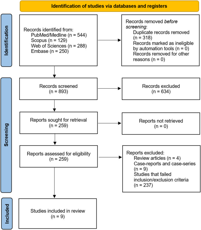
Methods: PubMed/MEDLINE, Web of Science, Scopus, and Embase were systematically searched up to August 27, 2024, to identify original English studies that compared OCT-A parameters between PwMS and PwNMOSD. The risk of bias across studies was evaluated utilizing the Newcastle-Ottawa Scale (NOS). Findings were consolidated using a narrative synthesis method.
Results: Nine studies involving 181 PwMS and 166 PwNMOSD were included. Compared to PwMS, PwNMOSD exhibited significantly lower vessel densities in the peripapillary and macular regions, reduced radial peripapillary capillary (RPC) density, and smaller foveal avascular zone (FAZ) areas, particularly in optic neuritis (ON)-affected eyes. Minimal differences were observed in eyes without ON, suggesting that ON may be crucial when utilizing OCT-A biomarkers for disease discrimination.
Conclusion: OCT-A metrics demonstrate potential as biomarkers that may help distinguish MS and NMOSD, with PwNMOSD showing more severe retinal vascular alterations. These preliminary findings highlight that OCT-A may hold promise as a diagnostic tool for differentiating MS and NMOSD. Further studies are needed to validate these findings.
期刊介绍:
International Journal of Retina and Vitreous focuses on the ophthalmic subspecialty of vitreoretinal disorders. The journal presents original articles on new approaches to diagnosis, outcomes of clinical trials, innovations in pharmacological therapy and surgical techniques, as well as basic science advances that impact clinical practice. Topical areas include, but are not limited to: -Imaging of the retina, choroid and vitreous -Innovations in optical coherence tomography (OCT) -Small-gauge vitrectomy, retinal detachment, chromovitrectomy -Electroretinography (ERG), microperimetry, other functional tests -Intraocular tumors -Retinal pharmacotherapy & drug delivery -Diabetic retinopathy & other vascular diseases -Age-related macular degeneration (AMD) & other macular entities

 求助内容:
求助内容: 应助结果提醒方式:


