Nancy Vargas, Andrés Quiroga, Juan Pablo Chaves, Harold Bolaños, ILKe Nalbantoglu, Claudia Patricia Acosta Astaiza, Yuefeng Wu, José B Sáenz
{"title":"Patterns of sialyl-Lewis X expression predict gastric histopathology.","authors":"Nancy Vargas, Andrés Quiroga, Juan Pablo Chaves, Harold Bolaños, ILKe Nalbantoglu, Claudia Patricia Acosta Astaiza, Yuefeng Wu, José B Sáenz","doi":"10.1186/s13000-025-01673-8","DOIUrl":null,"url":null,"abstract":"<p><strong>Introduction: </strong>Gastric cancer develops through a series of pre-cancerous changes over decades of chronic inflammation. Chronic atrophic gastritis (CAG) represents a critical transition in the progression to gastric cancer, though validated histologic markers are needed to more accurately detect and assess the extent of CAG. We previously identified sialyl-Lewis X (sLe<sup>x</sup>) as a marker of atrophic gastric epithelium in mice. Here, we establish patterns of sLe<sup>x</sup> expression that can be used to detect and distinguish human gastric pre-cancerous lesions.</p><p><strong>Methods: </strong>We obtained gastric corpus and/or antrum biopsies from 149 adult patients with dyspepsia. Biopsies were stained with hematoxylin/eosin and a commercially available antibody to sLe<sup>x</sup>. Histologic diagnoses included normal, chronic non-atrophic gastritis (CNG), or CAG with or without intestinal metaplasia (IM) and were determined by a single pathologist. A second pathologist graded each biopsy according to consensus criteria, based on the presence, intensity, and glandular distribution of sLe<sup>x</sup> staining. Log-linear models were used to determine the association between patterns of sLe<sup>x</sup> expression and gastric pathology.</p><p><strong>Results: </strong>The majority of patients (70%) had gastric pathology (CNG or CAG ± IM). The presence of sLe<sup>x</sup> could be used to detect gastric pathology (97% sensitivity), and the absence of sLe<sup>x</sup> staining could reliably predict normal histology (76% specificity). The intensity of sLe<sup>x</sup> staining significantly correlated with gastric pathology. Moreover, a deeper (≥ 50%) glandular sLe<sup>x</sup> distribution in the antrum was significantly associated with CAG, while a more superficial (< 50%) distribution significantly correlated with CNG.</p><p><strong>Conclusion: </strong>Patterns of sLe<sup>x</sup> expression can be used to detect and refine the histologic assessment of gastric pre-neoplastic lesion severity.</p>","PeriodicalId":11237,"journal":{"name":"Diagnostic Pathology","volume":"20 1","pages":"76"},"PeriodicalIF":2.3000,"publicationDate":"2025-06-23","publicationTypes":"Journal Article","fieldsOfStudy":null,"isOpenAccess":false,"openAccessPdf":"https://www.ncbi.nlm.nih.gov/pmc/articles/PMC12183862/pdf/","citationCount":"0","resultStr":null,"platform":"Semanticscholar","paperid":null,"PeriodicalName":"Diagnostic Pathology","FirstCategoryId":"3","ListUrlMain":"https://doi.org/10.1186/s13000-025-01673-8","RegionNum":3,"RegionCategory":"医学","ArticlePicture":[],"TitleCN":null,"AbstractTextCN":null,"PMCID":null,"EPubDate":"","PubModel":"","JCR":"Q2","JCRName":"PATHOLOGY","Score":null,"Total":0}
引用次数: 0
Abstract
Introduction: Gastric cancer develops through a series of pre-cancerous changes over decades of chronic inflammation. Chronic atrophic gastritis (CAG) represents a critical transition in the progression to gastric cancer, though validated histologic markers are needed to more accurately detect and assess the extent of CAG. We previously identified sialyl-Lewis X (sLex) as a marker of atrophic gastric epithelium in mice. Here, we establish patterns of sLex expression that can be used to detect and distinguish human gastric pre-cancerous lesions.
Methods: We obtained gastric corpus and/or antrum biopsies from 149 adult patients with dyspepsia. Biopsies were stained with hematoxylin/eosin and a commercially available antibody to sLex. Histologic diagnoses included normal, chronic non-atrophic gastritis (CNG), or CAG with or without intestinal metaplasia (IM) and were determined by a single pathologist. A second pathologist graded each biopsy according to consensus criteria, based on the presence, intensity, and glandular distribution of sLex staining. Log-linear models were used to determine the association between patterns of sLex expression and gastric pathology.
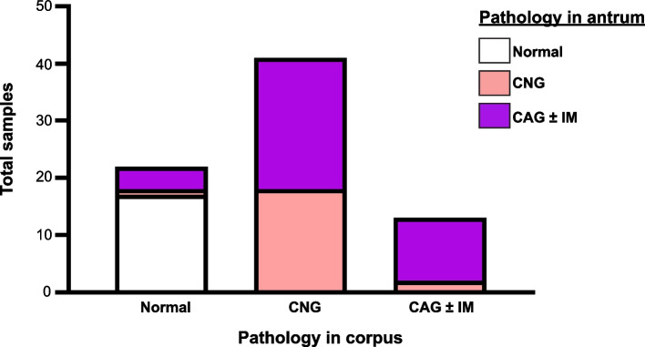
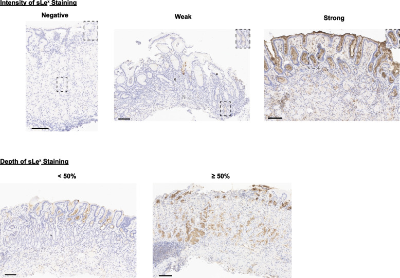
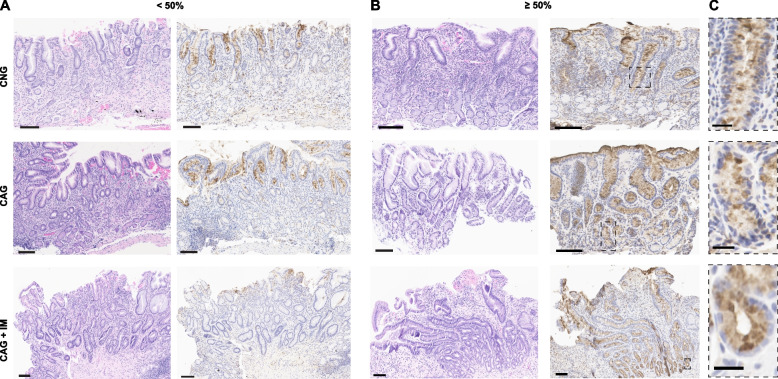
Results: The majority of patients (70%) had gastric pathology (CNG or CAG ± IM). The presence of sLex could be used to detect gastric pathology (97% sensitivity), and the absence of sLex staining could reliably predict normal histology (76% specificity). The intensity of sLex staining significantly correlated with gastric pathology. Moreover, a deeper (≥ 50%) glandular sLex distribution in the antrum was significantly associated with CAG, while a more superficial (< 50%) distribution significantly correlated with CNG.
Conclusion: Patterns of sLex expression can be used to detect and refine the histologic assessment of gastric pre-neoplastic lesion severity.
导言:胃癌是在几十年的慢性炎症中通过一系列癌前变化发展而来的。慢性萎缩性胃炎(CAG)是向胃癌发展的一个关键转变,尽管需要经过验证的组织学标志物来更准确地检测和评估CAG的程度。我们之前发现sialyl-Lewis X (sLex)是小鼠胃上皮萎缩的标记物。在这里,我们建立了可用于检测和区分人类胃癌前病变的sLex表达模式。方法:我们对149例成年消化不良患者进行了胃体和/或胃窦活检。活组织切片用苏木精/伊红染色和市售的sLex抗体染色。组织学诊断包括正常、慢性非萎缩性胃炎(CNG)、CAG伴或不伴肠化生(IM),并由一位病理学家确定。第二名病理学家根据一致的标准,根据sLex染色的存在、强度和腺体分布,对每个活检进行分级。使用对数线性模型来确定sLex表达模式与胃病理之间的关系。结果:绝大多数(70%)患者有胃病理(CNG或CAG±IM)。sLex的存在可以用于检测胃病理(97%的敏感性),而没有sLex染色可以可靠地预测正常组织学(76%的特异性)。sLex染色强度与胃病理有显著相关性。此外,在胃窦更深(≥50%)的腺体sLex分布与CAG显著相关,而更浅表的sLex表达模式可用于检测和完善胃癌前病变严重程度的组织学评估。
期刊介绍:
Diagnostic Pathology is an open access, peer-reviewed, online journal that considers research in surgical and clinical pathology, immunology, and biology, with a special focus on cutting-edge approaches in diagnostic pathology and tissue-based therapy. The journal covers all aspects of surgical pathology, including classic diagnostic pathology, prognosis-related diagnosis (tumor stages, prognosis markers, such as MIB-percentage, hormone receptors, etc.), and therapy-related findings. The journal also focuses on the technological aspects of pathology, including molecular biology techniques, morphometry aspects (stereology, DNA analysis, syntactic structure analysis), communication aspects (telecommunication, virtual microscopy, virtual pathology institutions, etc.), and electronic education and quality assurance (for example interactive publication, on-line references with automated updating, etc.).

 求助内容:
求助内容: 应助结果提醒方式:


