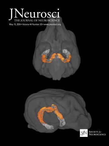
Diverse firing profiles of Crhbp-positive neurons in the dorsal pons suggestive of their pleiotropic roles in REM sleep regulation in mice.
IF 4.4
2区 医学
Q1 NEUROSCIENCES
引用次数: 0
Abstract
Rapid eye movement (REM) sleep is primarily regulated by the brainstem pons. In particular, the sublaterodorsal tegmentum (SubLDT) in the dorsal pons contains neurons whose activity is selective to REM sleep. Elucidation of the precise identities of these neurons and their roles in REM sleep regulation is challenging, however, due to the functional and molecular heterogeneity of the SubLDT. A recent study revealed that corticotropin-releasing hormone-binding protein (Crhbp)-positive neurons in the SubLDT projecting to the medulla play a crucial role in REM sleep regulation and that loss of these Crhbp-positive neurons underlies sleep deficits observed in Parkinson's disease. The firing patterns of these neurons during sleep/wake, however, remained unknown. Here, we used an opto-tagging method and conducted cell-type-specific recordings from Crhbp-positive neurons using a glass pipette microelectrode in unanesthetized male mice. We recorded 58 Crhbp-positive neurons and found that many of these neurons are REM sleep-active neurons (41.4%) and that the remaining neurons are mostly either wake-active, wake/REM sleep-active, or NREM sleep-active. In addition, projection-specific recordings revealed that the medulla-projecting Crhbp-positive neurons are mostly REM sleep-active neurons (75.0%). Based on clustering analysis and spike waveform analysis, REM sleep-active Crhbp-positive neurons can be further divided into different subtypes according to their electrophysiological properties, suggesting that Crhbp-positive neurons play diverse roles in REM sleep regulation.Significance statement Reduced REM sleep is a risk for dementia and mortality, suggesting it has critical roles in health. The mechanisms and functions of REM sleep, however, remain largely elusive. Classical electrophysiological studies identified neurons in the pons that are active during REM sleep, and a recent study revealed that Crhbp-positive neurons within the same area contribute to REM sleep regulation. The relationship between the neurons identified in each study, however, remained unknown. Loss of Crhbp-positive neurons underlies sleep deficits in Parkinson's disease, underscoring the importance of characterizing these neurons. Our study revealed that many of the Crhbp-positive neurons are REM sleep-active and comprise distinct subtypes in regard to firing patterns, suggesting their diverse roles in REM sleep regulation.脑桥背crhbp阳性神经元的不同放电谱提示其在小鼠快速眼动睡眠调节中的多效性作用。
快速眼动(REM)睡眠主要由脑干脑桥调节。特别是,脑桥背侧的嗅觉下被盖(subbldt)包含的神经元的活动对快速眼动睡眠有选择性。然而,由于subbldt的功能和分子异质性,阐明这些神经元的确切身份及其在REM睡眠调节中的作用具有挑战性。最近的一项研究表明,在投射到髓质的亚bldt中,促肾上腺皮质激素释放激素结合蛋白(Crhbp)阳性神经元在快速眼动睡眠调节中起着至关重要的作用,而这些Crhbp阳性神经元的缺失是帕金森病中观察到的睡眠缺陷的基础。然而,这些神经元在睡眠/清醒时的放电模式仍然未知。在这里,我们使用光标记方法,并使用玻璃移液管微电极对未麻醉的雄性小鼠的crhbp阳性神经元进行细胞类型特异性记录。我们记录了58个crhbp阳性神经元,发现这些神经元中有许多是REM睡眠活跃神经元(41.4%),其余的神经元大多是清醒活跃、清醒/REM睡眠活跃或NREM睡眠活跃。此外,投射特异性记录显示,髓质投射的crhbp阳性神经元主要是REM睡眠活跃神经元(75.0%)。基于聚类分析和尖峰波形分析,根据crhbp阳性神经元的电生理特性,可将REM睡眠活跃的crhbp阳性神经元进一步划分为不同的亚型,提示crhbp阳性神经元在REM睡眠调节中发挥着不同的作用。快速眼动睡眠减少会增加痴呆和死亡的风险,这表明它对健康有重要作用。然而,快速眼动睡眠的机制和功能在很大程度上仍然难以捉摸。经典的电生理学研究发现,脑桥上的神经元在快速眼动睡眠期间是活跃的,最近的一项研究表明,同一区域内的crhbp阳性神经元有助于快速眼动睡眠调节。然而,在每项研究中发现的神经元之间的关系仍然未知。crhbp阳性神经元的缺失是帕金森病睡眠不足的基础,这强调了表征这些神经元的重要性。我们的研究表明,许多crhbp阳性神经元是快速眼动睡眠活跃的,并且在放电模式方面包含不同的亚型,这表明它们在快速眼动睡眠调节中发挥着不同的作用。
本文章由计算机程序翻译,如有差异,请以英文原文为准。
求助全文
约1分钟内获得全文
求助全文
来源期刊

Journal of Neuroscience
医学-神经科学
CiteScore
9.30
自引率
3.80%
发文量
1164
审稿时长
12 months
期刊介绍:
JNeurosci (ISSN 0270-6474) is an official journal of the Society for Neuroscience. It is published weekly by the Society, fifty weeks a year, one volume a year. JNeurosci publishes papers on a broad range of topics of general interest to those working on the nervous system. Authors now have an Open Choice option for their published articles
 求助内容:
求助内容: 应助结果提醒方式:


