Ada Firrincieli, Eleonora Nardi, Lavinia Pugliese, Chiara Marconcini, Giovanni Alemanno, Luca Messerini
{"title":"Epidermoid Cyst of the Cecum Treated by Laparoscopic Colectomy: A Case Report With Histopathology and Literature Review.","authors":"Ada Firrincieli, Eleonora Nardi, Lavinia Pugliese, Chiara Marconcini, Giovanni Alemanno, Luca Messerini","doi":"10.1155/crgm/6326844","DOIUrl":null,"url":null,"abstract":"<p><p><b>Introduction:</b> Cecal epidermoid cyst (CEC) is a rare and benign lesion; the origin can be acquired or congenital, but the pathogenesis remains unclear. We present a case report of a patient with a cecal cyst treated by hemicolectomy. Histopathology revealed an epidermoid cyst (EC) of the cecum. <b>Case Presentation:</b> A 28-year-old woman was admitted to the hospital with abdominal pain, without significant past medical history. CT and MRI scans were performed, and a large cystic mass in the anterior portion of the pelvic region was detected. Imaging techniques managed to localize the site and dimensions of the neoplasm; however, they did not provide a conclusive diagnosis. The differential diagnosis was made with appendiceal mucocele, duplication cyst, or endometriotic cyst formation. Laparoscopic right hemicolectomy was performed; the mass did not present with any adhesions with the surrounding organs. Macroscopically, the mass appears as irregular extraluminal cystic lesion arising from the cecal wall of 104 × 83 × 68 mm. Microscopically, the cystic wall was lined by keratinized stratified squamous epithelium. No malignant findings were identified. Thus, the histopathologic evaluation leads to the final diagnosis of EC. <b>Conclusions:</b> ECs are rare benign neoplasms that can be acquired or congenital. They can vary both in their clinical and imaging presentation; the lesion can be associated with nonspecific symptoms or be asymptomatic. A wide heterogeneity both in sex distribution and age is observed. Imaging techniques are useful, but the final diagnosis can be made only after the complete surgical excision of the neoplasm and its histopathological examination.</p>","PeriodicalId":45645,"journal":{"name":"Case Reports in Gastrointestinal Medicine","volume":"2025 ","pages":"6326844"},"PeriodicalIF":0.5000,"publicationDate":"2025-06-13","publicationTypes":"Journal Article","fieldsOfStudy":null,"isOpenAccess":false,"openAccessPdf":"https://www.ncbi.nlm.nih.gov/pmc/articles/PMC12181667/pdf/","citationCount":"0","resultStr":null,"platform":"Semanticscholar","paperid":null,"PeriodicalName":"Case Reports in Gastrointestinal Medicine","FirstCategoryId":"1085","ListUrlMain":"https://doi.org/10.1155/crgm/6326844","RegionNum":0,"RegionCategory":null,"ArticlePicture":[],"TitleCN":null,"AbstractTextCN":null,"PMCID":null,"EPubDate":"2025/1/1 0:00:00","PubModel":"eCollection","JCR":"Q4","JCRName":"GASTROENTEROLOGY & HEPATOLOGY","Score":null,"Total":0}
引用次数: 0
Abstract
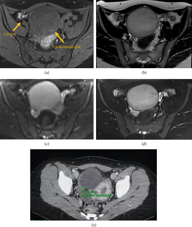
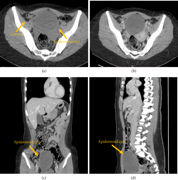
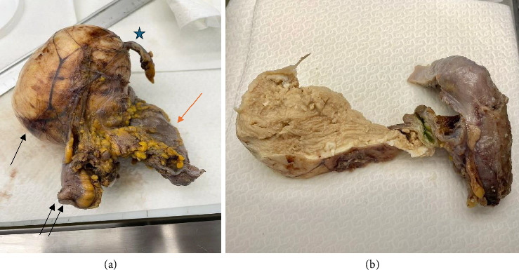
Introduction: Cecal epidermoid cyst (CEC) is a rare and benign lesion; the origin can be acquired or congenital, but the pathogenesis remains unclear. We present a case report of a patient with a cecal cyst treated by hemicolectomy. Histopathology revealed an epidermoid cyst (EC) of the cecum. Case Presentation: A 28-year-old woman was admitted to the hospital with abdominal pain, without significant past medical history. CT and MRI scans were performed, and a large cystic mass in the anterior portion of the pelvic region was detected. Imaging techniques managed to localize the site and dimensions of the neoplasm; however, they did not provide a conclusive diagnosis. The differential diagnosis was made with appendiceal mucocele, duplication cyst, or endometriotic cyst formation. Laparoscopic right hemicolectomy was performed; the mass did not present with any adhesions with the surrounding organs. Macroscopically, the mass appears as irregular extraluminal cystic lesion arising from the cecal wall of 104 × 83 × 68 mm. Microscopically, the cystic wall was lined by keratinized stratified squamous epithelium. No malignant findings were identified. Thus, the histopathologic evaluation leads to the final diagnosis of EC. Conclusions: ECs are rare benign neoplasms that can be acquired or congenital. They can vary both in their clinical and imaging presentation; the lesion can be associated with nonspecific symptoms or be asymptomatic. A wide heterogeneity both in sex distribution and age is observed. Imaging techniques are useful, but the final diagnosis can be made only after the complete surgical excision of the neoplasm and its histopathological examination.

 求助内容:
求助内容: 应助结果提醒方式:


