Robert W Gao, Judith As Jebastin, Doris E Wenger, William S Harmsen, Andrew L Folpe, Michael G Haddock, Ivy A Petersen, Safia K Ahmed
{"title":"Radiographic and pathologic response of myxoid liposarcoma treated with preoperative radiotherapy.","authors":"Robert W Gao, Judith As Jebastin, Doris E Wenger, William S Harmsen, Andrew L Folpe, Michael G Haddock, Ivy A Petersen, Safia K Ahmed","doi":"10.2478/raon-2025-0032","DOIUrl":null,"url":null,"abstract":"<p><strong>Background: </strong>We retrospectively assessed volumetric response of myxoid liposarcoma (MLPS) with preoperative radiotherapy (RT) and sought to identify disease and treatment characteristics associated with response.</p><p><strong>Patients and methods: </strong>We identified all patients with a histologic diagnosis of MLPS who received preoperative RT from 2013 to 2021 at our institution. We used cone beam computed tomography (CBCT) to assess changes in tumor volume and greatest dimension during treatment. Tumors were contoured on CBCT images prior to treatment and at the end of each week of RT. Percentage change in tumor volume and greatest dimension were calculated based on pre-treatment and final week contours. Patients with tumors incompletely visualized on CBCT were excluded from volume analysis but included on greatest dimension analysis. Magnetic resonance imaging (MRI) was used to evaluate pre- and post-RT radiographic features. Surgical pathology was reviewed to record pathologic characteristics.</p><p><strong>Results: </strong>Twenty patients met inclusion criteria. Most tumors (18/20) were low grade. The most frequent dose/fractionation scheme was 50 Gy in 25 fractions (16/20), with 3 patients receiving 36 Gy in 18 fractions. Median pre-RT volume and greatest dimension were 120 cc (interquartile range [IQR]: 56-399) and 11.2 cm (IQR: 8.4-14.1), respectively. Median percentage change in volume and greatest dimension were -37% (IQR: -57 to -29) and -10% (IQR: -20 to -7). All evaluable tumors decreased in volume during RT. Between pre- and post-RT MRI, most patients had a decrease in intratumoral (16/20) and peritumoral edema (11/20). Sixteen patients exhibited extensive pathologic response. There were no significant associations between radiographic and pathologic features and volumetric change. Local failure at 3 years was 9% (95% confidence interval: 1-59).</p><p><strong>Conclusions: </strong>We report significant decreases in MLPS tumor size during preoperative RT. There may be a role for adaptive RT planning to reduce target volumes and minimize RT-associated morbidity.</p>","PeriodicalId":21034,"journal":{"name":"Radiology and Oncology","volume":"59 2","pages":"176-182"},"PeriodicalIF":2.2000,"publicationDate":"2025-06-16","publicationTypes":"Journal Article","fieldsOfStudy":null,"isOpenAccess":false,"openAccessPdf":"https://www.ncbi.nlm.nih.gov/pmc/articles/PMC12182919/pdf/","citationCount":"0","resultStr":null,"platform":"Semanticscholar","paperid":null,"PeriodicalName":"Radiology and Oncology","FirstCategoryId":"3","ListUrlMain":"https://doi.org/10.2478/raon-2025-0032","RegionNum":4,"RegionCategory":"医学","ArticlePicture":[],"TitleCN":null,"AbstractTextCN":null,"PMCID":null,"EPubDate":"2025/6/1 0:00:00","PubModel":"eCollection","JCR":"Q3","JCRName":"ONCOLOGY","Score":null,"Total":0}
引用次数: 0
Abstract
Background: We retrospectively assessed volumetric response of myxoid liposarcoma (MLPS) with preoperative radiotherapy (RT) and sought to identify disease and treatment characteristics associated with response.
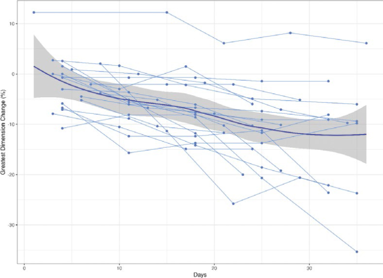
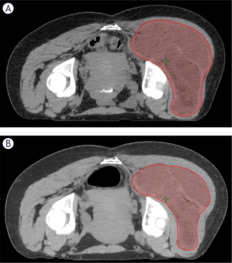
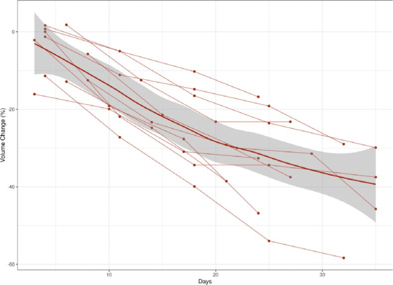
Patients and methods: We identified all patients with a histologic diagnosis of MLPS who received preoperative RT from 2013 to 2021 at our institution. We used cone beam computed tomography (CBCT) to assess changes in tumor volume and greatest dimension during treatment. Tumors were contoured on CBCT images prior to treatment and at the end of each week of RT. Percentage change in tumor volume and greatest dimension were calculated based on pre-treatment and final week contours. Patients with tumors incompletely visualized on CBCT were excluded from volume analysis but included on greatest dimension analysis. Magnetic resonance imaging (MRI) was used to evaluate pre- and post-RT radiographic features. Surgical pathology was reviewed to record pathologic characteristics.
Results: Twenty patients met inclusion criteria. Most tumors (18/20) were low grade. The most frequent dose/fractionation scheme was 50 Gy in 25 fractions (16/20), with 3 patients receiving 36 Gy in 18 fractions. Median pre-RT volume and greatest dimension were 120 cc (interquartile range [IQR]: 56-399) and 11.2 cm (IQR: 8.4-14.1), respectively. Median percentage change in volume and greatest dimension were -37% (IQR: -57 to -29) and -10% (IQR: -20 to -7). All evaluable tumors decreased in volume during RT. Between pre- and post-RT MRI, most patients had a decrease in intratumoral (16/20) and peritumoral edema (11/20). Sixteen patients exhibited extensive pathologic response. There were no significant associations between radiographic and pathologic features and volumetric change. Local failure at 3 years was 9% (95% confidence interval: 1-59).
Conclusions: We report significant decreases in MLPS tumor size during preoperative RT. There may be a role for adaptive RT planning to reduce target volumes and minimize RT-associated morbidity.
期刊介绍:
Radiology and Oncology is a multidisciplinary journal devoted to the publishing original and high quality scientific papers and review articles, pertinent to diagnostic and interventional radiology, computerized tomography, magnetic resonance, ultrasound, nuclear medicine, radiotherapy, clinical and experimental oncology, radiobiology, medical physics and radiation protection. Therefore, the scope of the journal is to cover beside radiology the diagnostic and therapeutic aspects in oncology, which distinguishes it from other journals in the field.
 求助内容:
求助内容: 应助结果提醒方式:


