{"title":"A Rare Presentation of Ruptured Pineal Region Teratoma with Postoperative Aseptic Meningitis.","authors":"Keisuke Fuji, Takumi Yamanaka, Manato Sakamoto, Ichita Taniyama, Yoshinobu Takahashi, Kazunori Tatsuzawa, Naoya Hashimoto","doi":"10.1159/000546099","DOIUrl":null,"url":null,"abstract":"<p><strong>Introduction: </strong>Mature teratomas are germ cell tumors composed of tissues derived from all three germ layers. These tumors are rare in the central nervous system, primarily occurring in the suprasellar and pineal regions. Rupture of intracranial teratomas is an exceptionally rare phenomenon, typically presenting on imaging as disseminated fatty droplets and occasionally associated with aseptic meningitis. We describe a case of a ruptured cystic teratoma in the pineal region, manifesting postoperatively with severe neurological symptoms consistent with aseptic meningitis.</p><p><strong>Case presentation: </strong>A 15-year-old boy presented with a 2-month history of persistent headaches. Computed tomography revealed a calcified mass lesion in the pineal region with low-density areas in the lateral ventricles. Magnetic resonance imaging (MRI) demonstrated a complex lesion in the pineal region and high T1 signal intensity bilaterally in the anterior horns of the lateral ventricles, suggestive of a ruptured teratoma. The patient underwent surgical resection. Postoperatively, he developed mild fever, severe headache, ocular pain, decreased vision, diplopia, and neck rigidity. Contrast-enhanced MRI revealed faint meningeal enhancement, consistent with aseptic meningitis. Symptoms gradually improved with steroid therapy.</p><p><strong>Conclusion: </strong>This case underscores the importance of recognizing rupture as a potential complication of intracranial teratomas, which may result in severe postoperative aseptic meningitis. Intraoperative measures, such as meticulous irrigation, are critical to mitigate this rare but serious complication.</p>","PeriodicalId":9625,"journal":{"name":"Case Reports in Oncology","volume":"18 1","pages":"817-823"},"PeriodicalIF":0.7000,"publicationDate":"2025-05-28","publicationTypes":"Journal Article","fieldsOfStudy":null,"isOpenAccess":false,"openAccessPdf":"https://www.ncbi.nlm.nih.gov/pmc/articles/PMC12180800/pdf/","citationCount":"0","resultStr":null,"platform":"Semanticscholar","paperid":null,"PeriodicalName":"Case Reports in Oncology","FirstCategoryId":"1085","ListUrlMain":"https://doi.org/10.1159/000546099","RegionNum":0,"RegionCategory":null,"ArticlePicture":[],"TitleCN":null,"AbstractTextCN":null,"PMCID":null,"EPubDate":"2025/1/1 0:00:00","PubModel":"eCollection","JCR":"Q4","JCRName":"ONCOLOGY","Score":null,"Total":0}
引用次数: 0
Abstract
Introduction: Mature teratomas are germ cell tumors composed of tissues derived from all three germ layers. These tumors are rare in the central nervous system, primarily occurring in the suprasellar and pineal regions. Rupture of intracranial teratomas is an exceptionally rare phenomenon, typically presenting on imaging as disseminated fatty droplets and occasionally associated with aseptic meningitis. We describe a case of a ruptured cystic teratoma in the pineal region, manifesting postoperatively with severe neurological symptoms consistent with aseptic meningitis.
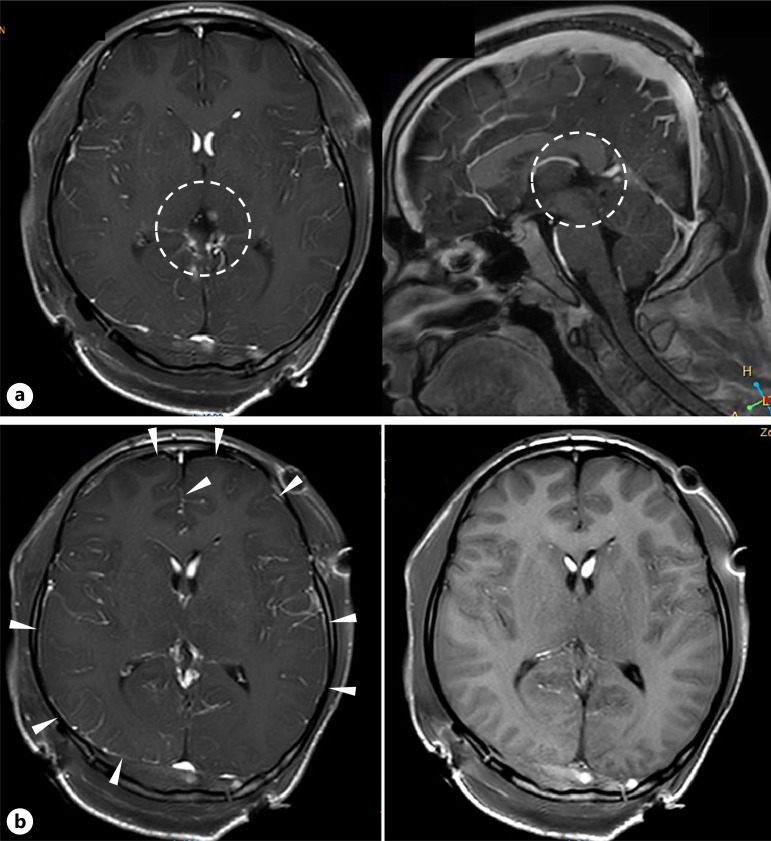
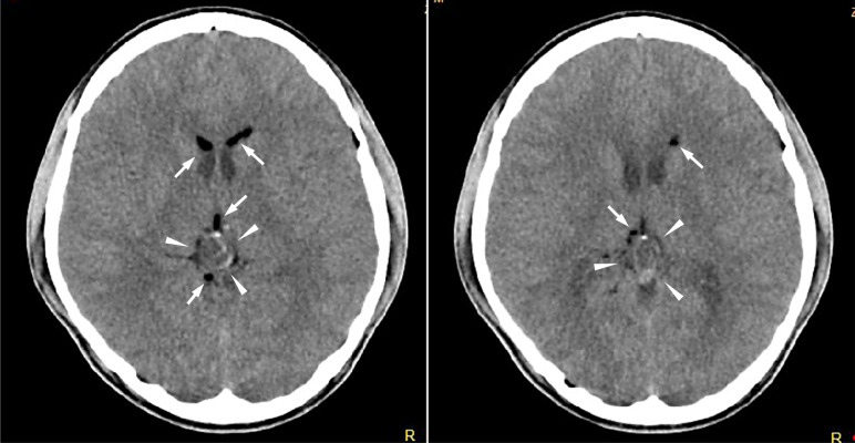
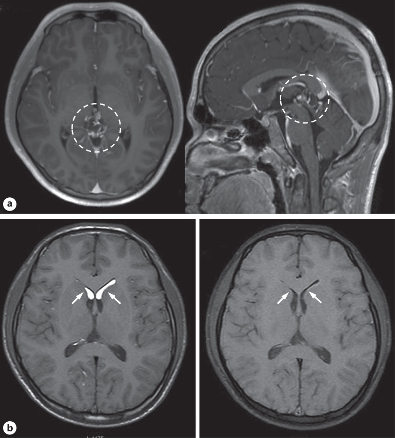
Case presentation: A 15-year-old boy presented with a 2-month history of persistent headaches. Computed tomography revealed a calcified mass lesion in the pineal region with low-density areas in the lateral ventricles. Magnetic resonance imaging (MRI) demonstrated a complex lesion in the pineal region and high T1 signal intensity bilaterally in the anterior horns of the lateral ventricles, suggestive of a ruptured teratoma. The patient underwent surgical resection. Postoperatively, he developed mild fever, severe headache, ocular pain, decreased vision, diplopia, and neck rigidity. Contrast-enhanced MRI revealed faint meningeal enhancement, consistent with aseptic meningitis. Symptoms gradually improved with steroid therapy.
Conclusion: This case underscores the importance of recognizing rupture as a potential complication of intracranial teratomas, which may result in severe postoperative aseptic meningitis. Intraoperative measures, such as meticulous irrigation, are critical to mitigate this rare but serious complication.

 求助内容:
求助内容: 应助结果提醒方式:


