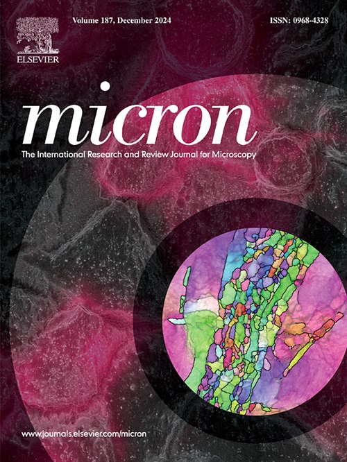
Identification of phase-separated structures and polymer crystals inside polyolefin blends
IF 2.2
3区 工程技术
Q1 MICROSCOPY
引用次数: 0
Abstract
Polyolefins, including polyethylene (PE) and isotactic polypropylene (iPP), constitute approximately half of all plastic production and consumption. However, recycled PE/iPP mixtures tend to be brittle, necessitating modifications to enhance their physical properties for practical applications. A comprehensive understanding of the internal structure of PE/iPP blends, which is influenced by phase separation and crystallization, is essential for improving their physical properties. Conventional electron microscopy techniques face limitations in distinguishing phases and observing crystal morphology. In this study, we introduce a four-dimensional scanning transmission electron microscopy technique known as nanodiffraction imaging (NDI) to analyze the internal structure of polyolefin blends. Scanning a 1.4 nm-diameter electron beam at 5 nm intervals, we examined a high-density PE (HDPE)/iPP blend specimen with a 10:90 wt composition. Based on scattering vectors and azimuthal angles of the diffraction peaks, NDI effectively identifies phases inside the HDPE/iPP blend, maps the spatial distribution of HDPE and iPP crystals, and facilitates the orientation mapping of their molecular chains. Additionally, it enables analysis of the relationship between HDPE and iPP crystals near the interface. The demonstrated effectiveness of NDI in structural analysis represents a significant advancement in understanding how the internal structure of polyolefin blends correlates with their physical properties.
聚烯烃共混物中相分离结构和聚合物晶体的鉴定
聚烯烃,包括聚乙烯(PE)和等规聚丙烯(iPP),约占所有塑料生产和消费的一半。然而,再生PE/iPP混合物往往是脆的,需要修改,以提高其实际应用的物理性能。全面了解PE/iPP共混物受相分离和结晶影响的内部结构,对于改善其物理性能至关重要。传统的电子显微镜技术在区分物相和观察晶体形态方面存在局限性。在这项研究中,我们引入了一种被称为纳米衍射成像(NDI)的四维扫描透射电子显微镜技术来分析聚烯烃共混物的内部结构。以5 nm的间隔扫描1.4 nm直径的电子束,我们检测了高密度PE (HDPE)/iPP混合样品,其重量比为10:90 。基于衍射峰的散射矢量和方位角,NDI可以有效地识别HDPE/iPP共混物内部的相,绘制HDPE和iPP晶体的空间分布,并便于分子链的取向作图。此外,它可以分析界面附近HDPE和iPP晶体之间的关系。NDI在结构分析中的有效性表明,在了解聚烯烃共混物的内部结构与其物理性质之间的关系方面取得了重大进展。
本文章由计算机程序翻译,如有差异,请以英文原文为准。
求助全文
约1分钟内获得全文
求助全文
来源期刊

Micron
工程技术-显微镜技术
CiteScore
4.30
自引率
4.20%
发文量
100
审稿时长
31 days
期刊介绍:
Micron is an interdisciplinary forum for all work that involves new applications of microscopy or where advanced microscopy plays a central role. The journal will publish on the design, methods, application, practice or theory of microscopy and microanalysis, including reports on optical, electron-beam, X-ray microtomography, and scanning-probe systems. It also aims at the regular publication of review papers, short communications, as well as thematic issues on contemporary developments in microscopy and microanalysis. The journal embraces original research in which microscopy has contributed significantly to knowledge in biology, life science, nanoscience and nanotechnology, materials science and engineering.
 求助内容:
求助内容: 应助结果提醒方式:


