{"title":"Diagnostic utility of cell block preparations from liquid-based cytology in cervical lesions: A comparative retrospective analysis.","authors":"Ceren Canbey, Sena Şen, Tevhide Bilgen Özcan","doi":"10.25259/Cytojournal_3_2025","DOIUrl":null,"url":null,"abstract":"<p><strong>Objective: </strong>Cervical cancer ranks as the fourth most prevalent cancer among women globally; it originates in the cervix and has a significant association with human papillomavirus (HPV) infection. The purpose of this study was to investigate the diagnostic utility of cell block (CB) preparations from liquid-based cytology samples in identifying cervical lesions among Turkish patients with HPV. This approach was intended to supplement conventional Pap smear tests and HPV testing.</p><p><strong>Material and methods: </strong>A retrospective analysis was conducted on 60 HPV-positive cervical smear samples processed through the ThinPrep Pap test. CBs were prepared from liquid-based residues, stained with hematoxylin and eosin, and analyzed. Cytological diagnoses were compared with histopathological findings from colposcopy-guided biopsies. The relationships between the Pap smear, CB, and biopsy results were statistically analyzed.</p><p><strong>Results: </strong>Pap smear cytology identified 1.6%, 16.6%, 43.3%, and 3.3% as high-grade squamous intraepithelial lesion (HSIL), low-grade squamous intraepithelial lesion (LSIL), atypical squamous cells of undetermined significance, and atypical squamous cells - HSIL cannot be excluded + LSIL, respectively. The CB evaluations classified 6.6% of the samples as cervical intraepithelial neoplasia (CIN)1, 1.6% as CIN2, and 1.6% as squamous cell carcinoma (SCC), with 78.3% deemed negative. Histopathological biopsy revealed CIN1 in 11.7%, CIN2 in 1.7%, and CIN3 in 8.3% of the patients. High concordance was observed between the Pap smear and CB diagnoses for negative and low-grade lesions, although discrepancies occurred in higher-grade lesions. HPV testing revealed 65% high-risk positivity, predominantly for HPV16 and HPV18. Significant correlations were found among HPV subtype positivity, CB, and biopsy diagnosis (<i>P</i> < 0.05).</p><p><strong>Conclusion: </strong>CB preparations provide enhanced diagnostic accuracy for high-grade lesions and SCC, thus complementing Pap smear cytology and HPV testing. This approach supports their integration into the routine cervical cancer screening protocols in Türkiye. Further global, multicenter studies are recommended to validate these findings.</p>","PeriodicalId":49082,"journal":{"name":"Cytojournal","volume":"22 ","pages":"48"},"PeriodicalIF":3.1000,"publicationDate":"2025-05-06","publicationTypes":"Journal Article","fieldsOfStudy":null,"isOpenAccess":false,"openAccessPdf":"https://www.ncbi.nlm.nih.gov/pmc/articles/PMC12178121/pdf/","citationCount":"0","resultStr":null,"platform":"Semanticscholar","paperid":null,"PeriodicalName":"Cytojournal","FirstCategoryId":"3","ListUrlMain":"https://doi.org/10.25259/Cytojournal_3_2025","RegionNum":4,"RegionCategory":"医学","ArticlePicture":[],"TitleCN":null,"AbstractTextCN":null,"PMCID":null,"EPubDate":"2025/1/1 0:00:00","PubModel":"eCollection","JCR":"Q2","JCRName":"PATHOLOGY","Score":null,"Total":0}
引用次数: 0
Abstract
Objective: Cervical cancer ranks as the fourth most prevalent cancer among women globally; it originates in the cervix and has a significant association with human papillomavirus (HPV) infection. The purpose of this study was to investigate the diagnostic utility of cell block (CB) preparations from liquid-based cytology samples in identifying cervical lesions among Turkish patients with HPV. This approach was intended to supplement conventional Pap smear tests and HPV testing.
Material and methods: A retrospective analysis was conducted on 60 HPV-positive cervical smear samples processed through the ThinPrep Pap test. CBs were prepared from liquid-based residues, stained with hematoxylin and eosin, and analyzed. Cytological diagnoses were compared with histopathological findings from colposcopy-guided biopsies. The relationships between the Pap smear, CB, and biopsy results were statistically analyzed.
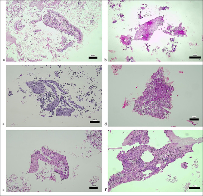
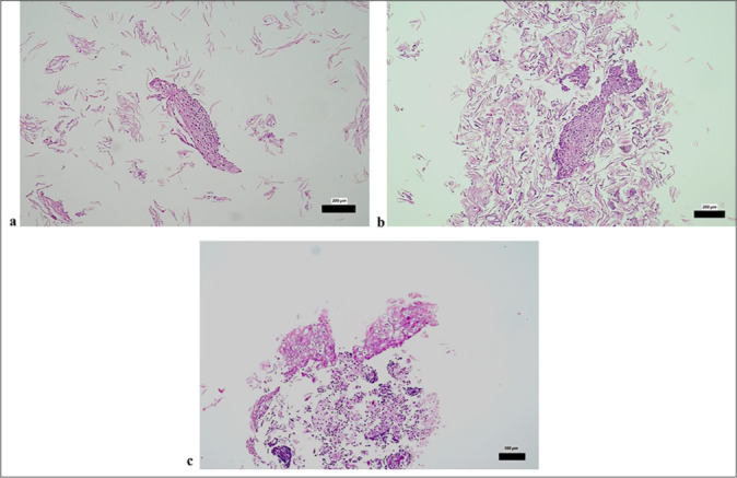
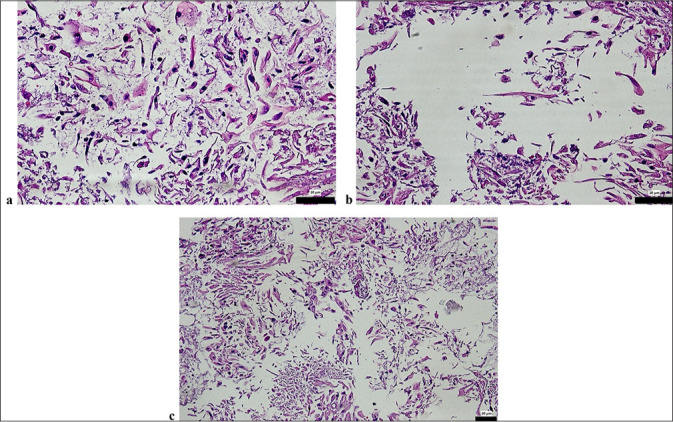
Results: Pap smear cytology identified 1.6%, 16.6%, 43.3%, and 3.3% as high-grade squamous intraepithelial lesion (HSIL), low-grade squamous intraepithelial lesion (LSIL), atypical squamous cells of undetermined significance, and atypical squamous cells - HSIL cannot be excluded + LSIL, respectively. The CB evaluations classified 6.6% of the samples as cervical intraepithelial neoplasia (CIN)1, 1.6% as CIN2, and 1.6% as squamous cell carcinoma (SCC), with 78.3% deemed negative. Histopathological biopsy revealed CIN1 in 11.7%, CIN2 in 1.7%, and CIN3 in 8.3% of the patients. High concordance was observed between the Pap smear and CB diagnoses for negative and low-grade lesions, although discrepancies occurred in higher-grade lesions. HPV testing revealed 65% high-risk positivity, predominantly for HPV16 and HPV18. Significant correlations were found among HPV subtype positivity, CB, and biopsy diagnosis (P < 0.05).
Conclusion: CB preparations provide enhanced diagnostic accuracy for high-grade lesions and SCC, thus complementing Pap smear cytology and HPV testing. This approach supports their integration into the routine cervical cancer screening protocols in Türkiye. Further global, multicenter studies are recommended to validate these findings.
期刊介绍:
The CytoJournal is an open-access peer-reviewed journal committed to publishing high-quality articles in the field of Diagnostic Cytopathology including Molecular aspects. The journal is owned by the Cytopathology Foundation and published by the Scientific Scholar.
 求助内容:
求助内容: 应助结果提醒方式:


