Robert E Ware, Damien Kee, Peter Roselt, Ivan Greguric, Andrew Katsifis, Thomas Bourdier, Wayne Noonan, William Murray, Catherine Mitchell, Marnie Downes, Mark Shackleton, Grant A McArthur, Rodney J Hicks
{"title":"Poor Diagnostic Performance of the Melanin-Binding Tracer [18 F]MEL050 in Human Melanoma Indicates Biological Heterogeneity.","authors":"Robert E Ware, Damien Kee, Peter Roselt, Ivan Greguric, Andrew Katsifis, Thomas Bourdier, Wayne Noonan, William Murray, Catherine Mitchell, Marnie Downes, Mark Shackleton, Grant A McArthur, Rodney J Hicks","doi":"10.1007/s11307-025-02025-0","DOIUrl":null,"url":null,"abstract":"<p><strong>Purpose: </strong>Malignant melanoma is a highly lethal malignancy typically characterized by the expression of melanin, which is an attractive diagnostic and therapeutic target in these cancers because it is expressed in few other tissues. Following preclinical evaluation of the melanin-targeting PET tracer, [18F]-6-fluoro-N-[2-(diethylamino)ethyl] pyridine-3-carboxamide ([18F]MEL050), we sought to evaluate this agent in patients with melanoma.</p><p><strong>Method: </strong>A phase I clinical trial was performed in ten patients with metastatic melanoma. Safety, dosimetry and diagnostic performance of intravenously administered][18F]MEL050 were evaluated. Based on results from this trial, we further assessed the prevalence and prognostic significance of loss of melanin expression in two historical patient cohorts for which there were matching histological and clinical outcome data.</p><p><strong>Results: </strong>Across the trial cohort, no adverse safety signals resulted from [18F]MEL050 administration. The whole-body effective dose was 0.0163 mSV/MBq for an adult male and 0.0206 mSV/MBq for an adult female. The human biodistribution was favorable with low uptake in organs at high risk of metastatic spread, including the brain. Of metastatic sites identified as melanoma on [18F]FDG PET/CT, only 31/65 (48%) were positive on [18F]MEL050 PET. Four [18F]FDG+[18F]MEL050+ metastases were resected from three patients and found to be melanotic by histological examination, whereas five [18F]FDG+[18F]MEL050- metastases from two patients were amelanotic. In our historical cohorts, amelanosis was more common in metastatic than primary disease (45% versus 20%) and the presence of melanin within sentinel lymph node metastases was associated with worse disease-free (HR 2.3 95% CI 1.3 - 4.3, p = 0.002) and disease-specific survivals (HR 3.6, 95% CI 1.4 - 9.7,p = 0.009) in stage III disease, compared with amelanotic sentinel lymph node metastases.</p><p><strong>Conclusion: </strong>We propose caution in the use of melanin-targeted agents for melanoma diagnosis and therapy until their utility as prognostic or predictive imaging biomarkers, and the biological implications of loss of melanin deposition during melanoma progression, are better understood.</p>","PeriodicalId":18760,"journal":{"name":"Molecular Imaging and Biology","volume":" ","pages":"649-657"},"PeriodicalIF":2.5000,"publicationDate":"2025-08-01","publicationTypes":"Journal Article","fieldsOfStudy":null,"isOpenAccess":false,"openAccessPdf":"https://www.ncbi.nlm.nih.gov/pmc/articles/PMC12405299/pdf/","citationCount":"0","resultStr":null,"platform":"Semanticscholar","paperid":null,"PeriodicalName":"Molecular Imaging and Biology","FirstCategoryId":"3","ListUrlMain":"https://doi.org/10.1007/s11307-025-02025-0","RegionNum":4,"RegionCategory":"医学","ArticlePicture":[],"TitleCN":null,"AbstractTextCN":null,"PMCID":null,"EPubDate":"2025/6/19 0:00:00","PubModel":"Epub","JCR":"Q2","JCRName":"RADIOLOGY, NUCLEAR MEDICINE & MEDICAL IMAGING","Score":null,"Total":0}
引用次数: 0
Abstract
Purpose: Malignant melanoma is a highly lethal malignancy typically characterized by the expression of melanin, which is an attractive diagnostic and therapeutic target in these cancers because it is expressed in few other tissues. Following preclinical evaluation of the melanin-targeting PET tracer, [18F]-6-fluoro-N-[2-(diethylamino)ethyl] pyridine-3-carboxamide ([18F]MEL050), we sought to evaluate this agent in patients with melanoma.
Method: A phase I clinical trial was performed in ten patients with metastatic melanoma. Safety, dosimetry and diagnostic performance of intravenously administered][18F]MEL050 were evaluated. Based on results from this trial, we further assessed the prevalence and prognostic significance of loss of melanin expression in two historical patient cohorts for which there were matching histological and clinical outcome data.
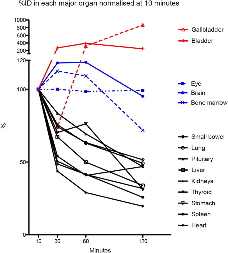
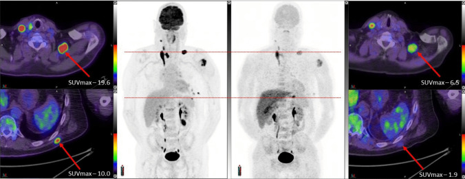
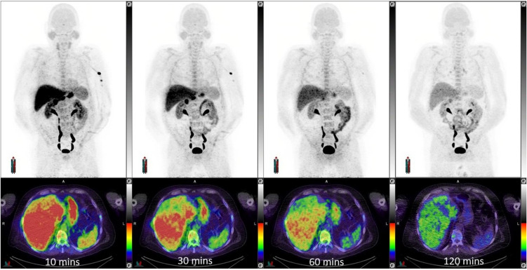
Results: Across the trial cohort, no adverse safety signals resulted from [18F]MEL050 administration. The whole-body effective dose was 0.0163 mSV/MBq for an adult male and 0.0206 mSV/MBq for an adult female. The human biodistribution was favorable with low uptake in organs at high risk of metastatic spread, including the brain. Of metastatic sites identified as melanoma on [18F]FDG PET/CT, only 31/65 (48%) were positive on [18F]MEL050 PET. Four [18F]FDG+[18F]MEL050+ metastases were resected from three patients and found to be melanotic by histological examination, whereas five [18F]FDG+[18F]MEL050- metastases from two patients were amelanotic. In our historical cohorts, amelanosis was more common in metastatic than primary disease (45% versus 20%) and the presence of melanin within sentinel lymph node metastases was associated with worse disease-free (HR 2.3 95% CI 1.3 - 4.3, p = 0.002) and disease-specific survivals (HR 3.6, 95% CI 1.4 - 9.7,p = 0.009) in stage III disease, compared with amelanotic sentinel lymph node metastases.
Conclusion: We propose caution in the use of melanin-targeted agents for melanoma diagnosis and therapy until their utility as prognostic or predictive imaging biomarkers, and the biological implications of loss of melanin deposition during melanoma progression, are better understood.
期刊介绍:
Molecular Imaging and Biology (MIB) invites original contributions (research articles, review articles, commentaries, etc.) on the utilization of molecular imaging (i.e., nuclear imaging, optical imaging, autoradiography and pathology, MRI, MPI, ultrasound imaging, radiomics/genomics etc.) to investigate questions related to biology and health. The objective of MIB is to provide a forum to the discovery of molecular mechanisms of disease through the use of imaging techniques. We aim to investigate the biological nature of disease in patients and establish new molecular imaging diagnostic and therapy procedures.
Some areas that are covered are:
Preclinical and clinical imaging of macromolecular targets (e.g., genes, receptors, enzymes) involved in significant biological processes.
The design, characterization, and study of new molecular imaging probes and contrast agents for the functional interrogation of macromolecular targets.
Development and evaluation of imaging systems including instrumentation, image reconstruction algorithms, image analysis, and display.
Development of molecular assay approaches leading to quantification of the biological information obtained in molecular imaging.
Study of in vivo animal models of disease for the development of new molecular diagnostics and therapeutics.
Extension of in vitro and in vivo discoveries using disease models, into well designed clinical research investigations.
Clinical molecular imaging involving clinical investigations, clinical trials and medical management or cost-effectiveness studies.

 求助内容:
求助内容: 应助结果提醒方式:


