Early stage prediction of bone regeneration using FEA and cell differentiation algorithms with 3D-printed PLA and PCL scaffolds: modeling and application to dorsal double-plating in distal radius fractures.
IF 3.1 Q1 RADIOLOGY, NUCLEAR MEDICINE & MEDICAL IMAGING
Hsuan Chih Liu, Ya-Han Chan, Shao-Fu Huang, Wei-Che Tsai, Yen Cheng, Chun-Li Lin
{"title":"Early stage prediction of bone regeneration using FEA and cell differentiation algorithms with 3D-printed PLA and PCL scaffolds: modeling and application to dorsal double-plating in distal radius fractures.","authors":"Hsuan Chih Liu, Ya-Han Chan, Shao-Fu Huang, Wei-Che Tsai, Yen Cheng, Chun-Li Lin","doi":"10.1186/s41205-025-00278-7","DOIUrl":null,"url":null,"abstract":"<p><p>This study introduces an advanced framework that integrates biphasic cell differentiation bone remodeling theory with finite element (FE) analysis and multi-remodeling simulation to evaluate the performance of 3D-printed biodegradable scaffolds for bone defect repair. The program incorporates a time-dependent cell differentiation stimulus (S), accounting for fluid-phase shear stress and solid-phase shear strain, to dynamically predict bone cell behavior. The study focuses on polylactic acid (PLA) and polycaprolactone (PCL) scaffolds with diamond (DU) and random (YM) lattice designs, applied to a dorsal double-plating (DDP) fixation model for distal radius fractures. Material testing reveals that PLA provides higher rigidity and strength, while PCL offers superior ductility. Mechanical strength tests confirm the superior performance of DU lattice structures under compression, shear, and torsion forces. The bone remodeling program, applied to 36 model combinations of fracture gaps, materials, and lattice designs, computes the total percentage of cell differentiation (TPCD), identifying scaffold material as the key factor, with PLA significantly enhancing TPCD values. Biomechanical analysis after 50 remodeling iterations in a 5.4 mm fracture gap shows that the PLA + DU scaffold reduces displacement by 35%/39%/75%, bone stress by 19%/16%/67%, and fixation plate stress by 77%/66%/93% under axial/bending/torsion loads, respectively, compared to the PCL + YM scaffold. This study highlights the critical role of dynamic remodeling programs in optimizing scaffold material properties and lattice architectures, establishing a robust platform for patient-specific bone repair solutions in regenerative medicine.</p>","PeriodicalId":72036,"journal":{"name":"3D printing in medicine","volume":"11 1","pages":"30"},"PeriodicalIF":3.1000,"publicationDate":"2025-06-19","publicationTypes":"Journal Article","fieldsOfStudy":null,"isOpenAccess":false,"openAccessPdf":"https://www.ncbi.nlm.nih.gov/pmc/articles/PMC12177956/pdf/","citationCount":"0","resultStr":null,"platform":"Semanticscholar","paperid":null,"PeriodicalName":"3D printing in medicine","FirstCategoryId":"1085","ListUrlMain":"https://doi.org/10.1186/s41205-025-00278-7","RegionNum":0,"RegionCategory":null,"ArticlePicture":[],"TitleCN":null,"AbstractTextCN":null,"PMCID":null,"EPubDate":"","PubModel":"","JCR":"Q1","JCRName":"RADIOLOGY, NUCLEAR MEDICINE & MEDICAL IMAGING","Score":null,"Total":0}
引用次数: 0
Abstract
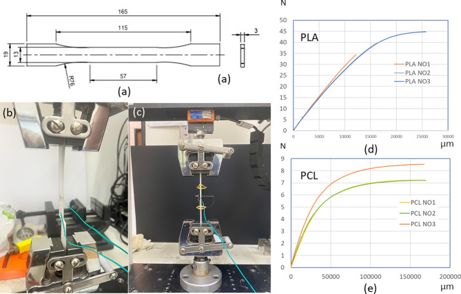
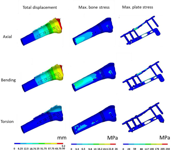
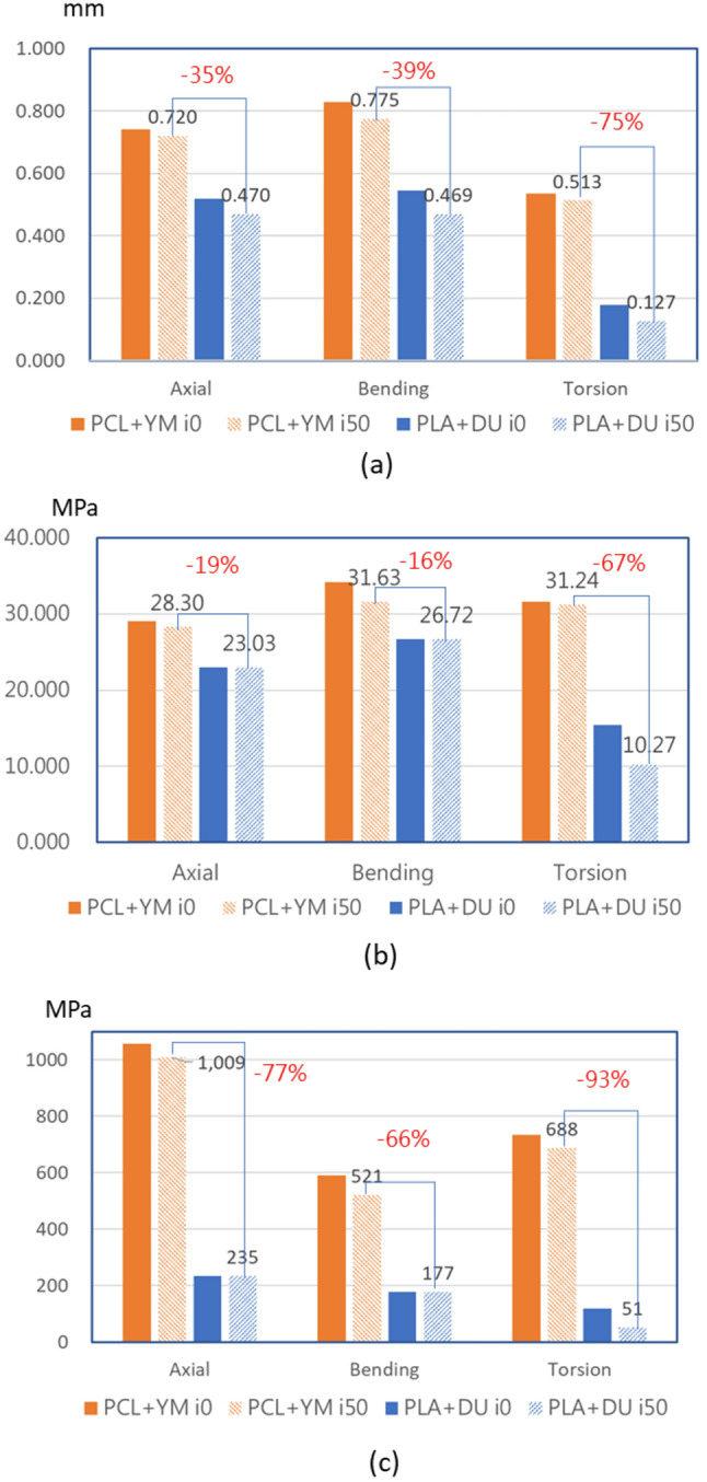
This study introduces an advanced framework that integrates biphasic cell differentiation bone remodeling theory with finite element (FE) analysis and multi-remodeling simulation to evaluate the performance of 3D-printed biodegradable scaffolds for bone defect repair. The program incorporates a time-dependent cell differentiation stimulus (S), accounting for fluid-phase shear stress and solid-phase shear strain, to dynamically predict bone cell behavior. The study focuses on polylactic acid (PLA) and polycaprolactone (PCL) scaffolds with diamond (DU) and random (YM) lattice designs, applied to a dorsal double-plating (DDP) fixation model for distal radius fractures. Material testing reveals that PLA provides higher rigidity and strength, while PCL offers superior ductility. Mechanical strength tests confirm the superior performance of DU lattice structures under compression, shear, and torsion forces. The bone remodeling program, applied to 36 model combinations of fracture gaps, materials, and lattice designs, computes the total percentage of cell differentiation (TPCD), identifying scaffold material as the key factor, with PLA significantly enhancing TPCD values. Biomechanical analysis after 50 remodeling iterations in a 5.4 mm fracture gap shows that the PLA + DU scaffold reduces displacement by 35%/39%/75%, bone stress by 19%/16%/67%, and fixation plate stress by 77%/66%/93% under axial/bending/torsion loads, respectively, compared to the PCL + YM scaffold. This study highlights the critical role of dynamic remodeling programs in optimizing scaffold material properties and lattice architectures, establishing a robust platform for patient-specific bone repair solutions in regenerative medicine.

 求助内容:
求助内容: 应助结果提醒方式:


