{"title":"Choroidal changes after vitrectomy performed for macular hole retinal detachment.","authors":"Hirotaka Sumida, Ikuko Umeda, Takayuki Baba","doi":"10.1007/s10384-025-01218-y","DOIUrl":null,"url":null,"abstract":"<p><strong>Purpose: </strong>To investigate changes in the choroidal area (CA), luminal area (LA), stromal area (SA), choroidal vascularity index (CVI), and choroidal thickness (CT) before and after vitrectomy in eyes with macular hole retinal detachment (MHRD).</p><p><strong>Study design: </strong>Retrospective observational study.</p><p><strong>Methods: </strong>We measured the best-corrected visual acuity (BCVA), intraocular pressure (IOP), CA, LA, SA, CVI, and CT preoperatively and 1, 3, and 6 months postoperatively in 10 eyes with MHRD. CA was measured within a 3-mm-wide area around the fovea in the horizontal and vertical images. LA and SA were quantified using the Niblack method, and CVI was calculated as the ratio of LA to CA. CT was measured at the subfovea and at 1 and 3 mm vertically and horizontally away from the fovea.</p><p><strong>Results: </strong>BCVA improved significantly at 1 and 3 months postoperatively (P = 0.036 and 0.016). IOP remained stable. CA and LA decreased significantly 6 months postoperatively in both the horizontal (P = 0.002 and 0.014) and vertical sections (P = 0.006 and 0.002). SA remained stable. CVI reduced significantly at 1 month horizontally and at 3 months vertically (both P = 0.027). CT decreased significantly in the subfovea at 3 and 6 months postoperatively (P = 0.027 and 0.020, respectively). Significant reductions were also observed at 1 mm nasal, temporal, and superior regions (P = 0.014, 0.014, and 0.004) and at 2 mm temporal and superior regions 1 month postoperatively (P = 0.020 and 0.014).</p><p><strong>Conclusion: </strong>Choroidal thinning was observed after vitrectomy in eyes with MHRD, driven by a reduction in the luminal area.</p>","PeriodicalId":14563,"journal":{"name":"Japanese Journal of Ophthalmology","volume":" ","pages":"755-765"},"PeriodicalIF":1.9000,"publicationDate":"2025-09-01","publicationTypes":"Journal Article","fieldsOfStudy":null,"isOpenAccess":false,"openAccessPdf":"https://www.ncbi.nlm.nih.gov/pmc/articles/PMC12390876/pdf/","citationCount":"0","resultStr":null,"platform":"Semanticscholar","paperid":null,"PeriodicalName":"Japanese Journal of Ophthalmology","FirstCategoryId":"3","ListUrlMain":"https://doi.org/10.1007/s10384-025-01218-y","RegionNum":3,"RegionCategory":"医学","ArticlePicture":[],"TitleCN":null,"AbstractTextCN":null,"PMCID":null,"EPubDate":"2025/6/18 0:00:00","PubModel":"Epub","JCR":"Q2","JCRName":"OPHTHALMOLOGY","Score":null,"Total":0}
引用次数: 0
Abstract
Purpose: To investigate changes in the choroidal area (CA), luminal area (LA), stromal area (SA), choroidal vascularity index (CVI), and choroidal thickness (CT) before and after vitrectomy in eyes with macular hole retinal detachment (MHRD).
Study design: Retrospective observational study.
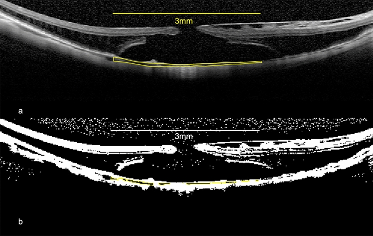
Methods: We measured the best-corrected visual acuity (BCVA), intraocular pressure (IOP), CA, LA, SA, CVI, and CT preoperatively and 1, 3, and 6 months postoperatively in 10 eyes with MHRD. CA was measured within a 3-mm-wide area around the fovea in the horizontal and vertical images. LA and SA were quantified using the Niblack method, and CVI was calculated as the ratio of LA to CA. CT was measured at the subfovea and at 1 and 3 mm vertically and horizontally away from the fovea.
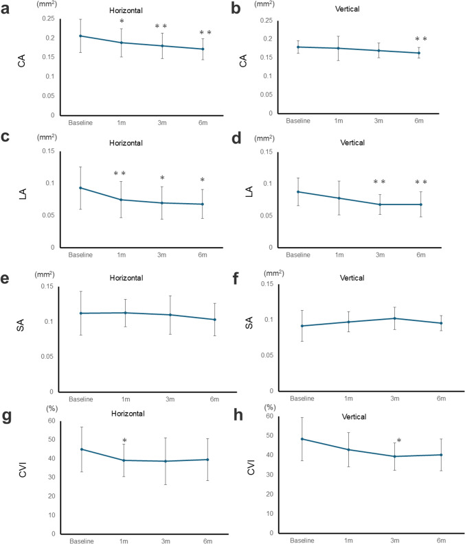
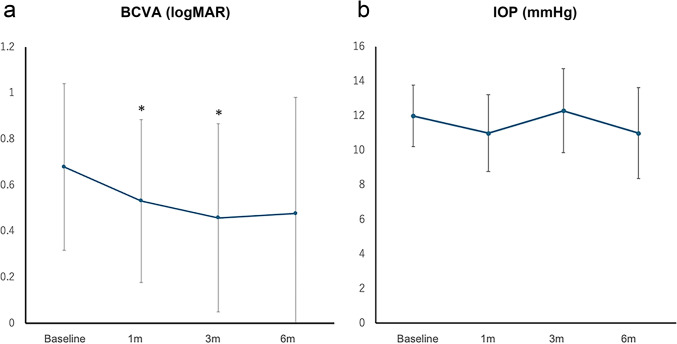
Results: BCVA improved significantly at 1 and 3 months postoperatively (P = 0.036 and 0.016). IOP remained stable. CA and LA decreased significantly 6 months postoperatively in both the horizontal (P = 0.002 and 0.014) and vertical sections (P = 0.006 and 0.002). SA remained stable. CVI reduced significantly at 1 month horizontally and at 3 months vertically (both P = 0.027). CT decreased significantly in the subfovea at 3 and 6 months postoperatively (P = 0.027 and 0.020, respectively). Significant reductions were also observed at 1 mm nasal, temporal, and superior regions (P = 0.014, 0.014, and 0.004) and at 2 mm temporal and superior regions 1 month postoperatively (P = 0.020 and 0.014).
Conclusion: Choroidal thinning was observed after vitrectomy in eyes with MHRD, driven by a reduction in the luminal area.
期刊介绍:
The Japanese Journal of Ophthalmology (JJO) was inaugurated in 1957 as a quarterly journal published in English by the Ophthalmology Department of the University of Tokyo, with the aim of disseminating the achievements of Japanese ophthalmologists worldwide. JJO remains the only Japanese ophthalmology journal published in English. In 1997, the Japanese Ophthalmological Society assumed the responsibility for publishing the Japanese Journal of Ophthalmology as its official English-language publication.
Currently the journal is published bimonthly and accepts papers from authors worldwide. JJO has become an international interdisciplinary forum for the publication of basic science and clinical research papers.

 求助内容:
求助内容: 应助结果提醒方式:


