Rachael Loek, David Gardiner, George Moore, Ashfaq Marghoob, Carine Laporte
{"title":"Dermoscopic evaluation of normal canine skin with a handheld dermoscope.","authors":"Rachael Loek, David Gardiner, George Moore, Ashfaq Marghoob, Carine Laporte","doi":"10.1111/vde.13368","DOIUrl":null,"url":null,"abstract":"<p><strong>Background: </strong>Dermoscopy is a noninvasive diagnostic tool that provides a magnified view of skin structures. While dermoscopy is described for certain canine dermatological diseases, large-scale studies evaluating normal skin are lacking.</p><p><strong>Hypothesis/objective: </strong>This study aimed to correlate dermoscopic findings with histopathological results in healthy canine skin to enhance understanding of dermoscopic microanatomy and pigmentation patterns.</p><p><strong>Animals: </strong>Healthy, adult, shelter dogs (n = 121).</p><p><strong>Materials and methods: </strong>After general anaesthesia for prescheduled sterilisation procedures, four regions on each dog were assessed using a handheld dermoscope followed by collecting a biopsy for histopathological investigation. Dermoscopic assessment included skin colour and pattern, presence of scale and blood vessel number. Dermoscopic findings were correlated with histopathological characteristics.</p><p><strong>Results: </strong>Dermoscopy identified grey as the most common skin colour, diffuse as the primary pattern, most commonly mild scale and primarily absent blood vessels. Dermoscopy correlation with histopathological results identified moderate scale as more likely to have hyperkeratosis, and no significant correlation between visualised blood vessels and number of endothelial cells. Furthermore, the dermoscopic colour brown was more likely to have melanin within each epidermal layer, while white was less likely to have melanin within each layer. Despite the lack of gross and dermoscopic inflammation, such as erythema, 53 of 484 sites had histopathological evidence of inflammation, with primarily mild mastocytic and eosinophilic superficial dermatitis.</p><p><strong>Conclusions and clinical relevance: </strong>Dermoscopy can identify characteristics in canine skin that correlate with histopathological results, yet mild inflammation may remain undetected. This correlation better establishes baselines for future studies utilising dermoscopy when assessing dermatological diseases.</p>","PeriodicalId":23599,"journal":{"name":"Veterinary dermatology","volume":" ","pages":"593-601"},"PeriodicalIF":1.4000,"publicationDate":"2025-10-01","publicationTypes":"Journal Article","fieldsOfStudy":null,"isOpenAccess":false,"openAccessPdf":"https://www.ncbi.nlm.nih.gov/pmc/articles/PMC12420875/pdf/","citationCount":"0","resultStr":null,"platform":"Semanticscholar","paperid":null,"PeriodicalName":"Veterinary dermatology","FirstCategoryId":"97","ListUrlMain":"https://doi.org/10.1111/vde.13368","RegionNum":3,"RegionCategory":"农林科学","ArticlePicture":[],"TitleCN":null,"AbstractTextCN":null,"PMCID":null,"EPubDate":"2025/6/17 0:00:00","PubModel":"Epub","JCR":"Q3","JCRName":"DERMATOLOGY","Score":null,"Total":0}
引用次数: 0
Abstract
Background: Dermoscopy is a noninvasive diagnostic tool that provides a magnified view of skin structures. While dermoscopy is described for certain canine dermatological diseases, large-scale studies evaluating normal skin are lacking.
Hypothesis/objective: This study aimed to correlate dermoscopic findings with histopathological results in healthy canine skin to enhance understanding of dermoscopic microanatomy and pigmentation patterns.
Animals: Healthy, adult, shelter dogs (n = 121).
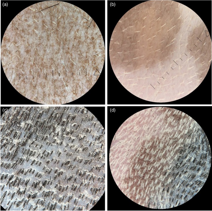
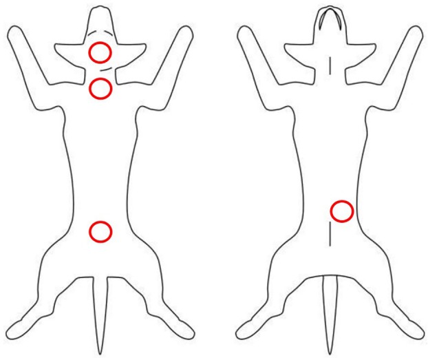
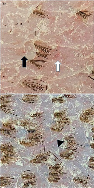
Materials and methods: After general anaesthesia for prescheduled sterilisation procedures, four regions on each dog were assessed using a handheld dermoscope followed by collecting a biopsy for histopathological investigation. Dermoscopic assessment included skin colour and pattern, presence of scale and blood vessel number. Dermoscopic findings were correlated with histopathological characteristics.
Results: Dermoscopy identified grey as the most common skin colour, diffuse as the primary pattern, most commonly mild scale and primarily absent blood vessels. Dermoscopy correlation with histopathological results identified moderate scale as more likely to have hyperkeratosis, and no significant correlation between visualised blood vessels and number of endothelial cells. Furthermore, the dermoscopic colour brown was more likely to have melanin within each epidermal layer, while white was less likely to have melanin within each layer. Despite the lack of gross and dermoscopic inflammation, such as erythema, 53 of 484 sites had histopathological evidence of inflammation, with primarily mild mastocytic and eosinophilic superficial dermatitis.
Conclusions and clinical relevance: Dermoscopy can identify characteristics in canine skin that correlate with histopathological results, yet mild inflammation may remain undetected. This correlation better establishes baselines for future studies utilising dermoscopy when assessing dermatological diseases.
期刊介绍:
Veterinary Dermatology is a bi-monthly, peer-reviewed, international journal which publishes papers on all aspects of the skin of mammals, birds, reptiles, amphibians and fish. Scientific research papers, clinical case reports and reviews covering the following aspects of dermatology will be considered for publication:
-Skin structure (anatomy, histology, ultrastructure)
-Skin function (physiology, biochemistry, pharmacology, immunology, genetics)
-Skin microbiology and parasitology
-Dermatopathology
-Pathogenesis, diagnosis and treatment of skin diseases
-New disease entities
 求助内容:
求助内容: 应助结果提醒方式:


