Shuoqi Zhang, Jiayi Ma, Shaolong Wu, Ziwei Hu, Su Yan, Junghun Cho, Yi Wang, Lingli Dong, Shun Zhang, Wenzhen Zhu
{"title":"Evaluation of MRI-based brain oxygen extraction fraction mapping in patients with systemic lupus erythematosus.","authors":"Shuoqi Zhang, Jiayi Ma, Shaolong Wu, Ziwei Hu, Su Yan, Junghun Cho, Yi Wang, Lingli Dong, Shun Zhang, Wenzhen Zhu","doi":"10.1136/lupus-2025-001522","DOIUrl":null,"url":null,"abstract":"<p><strong>Objective: </strong>We aim to assess the cerebral oxygen extraction fraction (OEF) changes in patients with systemic lupus erythematosus (SLE) and neuropsychiatric SLE (NPSLE) by using an MRI-based technique and examine the relationship between OEF and cognition.</p><p><strong>Methods: </strong>43 SLE patients (18 NPSLE and 25 non-NPSLE) and 26 healthy controls (HC) were recruited. Cognitive function was assessed via the Montreal Cognitive Assessment (MoCA). OEF was calculated by quantitative susceptibility mapping plus quantitative blood oxygen level-dependent model (QQ). Whole-brain voxel-wise analysis of OEF was performed. In subcortical grey matter structures, regional OEF values were measured, and their relationship with MoCA scores was explored.</p><p><strong>Results: </strong>Whole-brain voxel-wise analysis revealed significant changes of OEF primarily in the limbic system, including the orbitofrontal cortex and bilateral insular lobes, among HC, non-NPSLE and NPSLE groups. Regional analysis indicated reduced OEF values in subregions of the amygdala, hippocampus and caudate nucleus in non-NPSLE compared with HC, with decreasing trends observed in all selected regions of subcortical grey matter structures. In the right hippocampus, OEF values were increased in NPSLE patients compared with non-NPSLE patients. Considering all subjects in the study, OEF values in the bilateral medial amygdalae, right lateral amygdala, left rostral hippocampus and right dorsal caudate nucleus were positively correlated with MoCA scores.</p><p><strong>Conclusion: </strong>Cerebral OEF mapping in patients with SLE is readily available using the MRI-based QQ method, which has the potential to serve as an adjunctive tool for diagnosing NPSLE and monitoring cognitive impairment in SLE.</p>","PeriodicalId":18126,"journal":{"name":"Lupus Science & Medicine","volume":"12 1","pages":""},"PeriodicalIF":3.5000,"publicationDate":"2025-06-16","publicationTypes":"Journal Article","fieldsOfStudy":null,"isOpenAccess":false,"openAccessPdf":"https://www.ncbi.nlm.nih.gov/pmc/articles/PMC12182206/pdf/","citationCount":"0","resultStr":null,"platform":"Semanticscholar","paperid":null,"PeriodicalName":"Lupus Science & Medicine","FirstCategoryId":"3","ListUrlMain":"https://doi.org/10.1136/lupus-2025-001522","RegionNum":2,"RegionCategory":"医学","ArticlePicture":[],"TitleCN":null,"AbstractTextCN":null,"PMCID":null,"EPubDate":"","PubModel":"","JCR":"Q1","JCRName":"RHEUMATOLOGY","Score":null,"Total":0}
引用次数: 0
Abstract
Objective: We aim to assess the cerebral oxygen extraction fraction (OEF) changes in patients with systemic lupus erythematosus (SLE) and neuropsychiatric SLE (NPSLE) by using an MRI-based technique and examine the relationship between OEF and cognition.
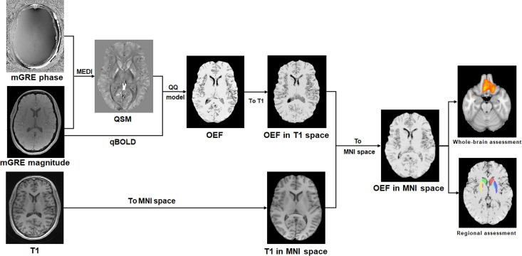
Methods: 43 SLE patients (18 NPSLE and 25 non-NPSLE) and 26 healthy controls (HC) were recruited. Cognitive function was assessed via the Montreal Cognitive Assessment (MoCA). OEF was calculated by quantitative susceptibility mapping plus quantitative blood oxygen level-dependent model (QQ). Whole-brain voxel-wise analysis of OEF was performed. In subcortical grey matter structures, regional OEF values were measured, and their relationship with MoCA scores was explored.
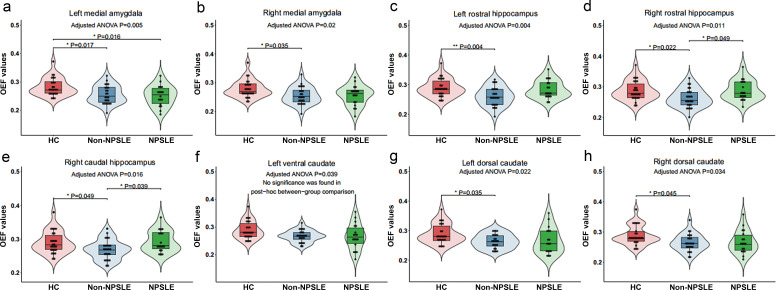
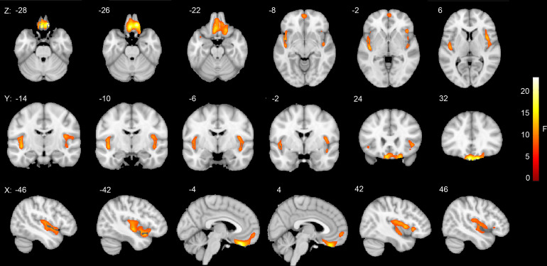
Results: Whole-brain voxel-wise analysis revealed significant changes of OEF primarily in the limbic system, including the orbitofrontal cortex and bilateral insular lobes, among HC, non-NPSLE and NPSLE groups. Regional analysis indicated reduced OEF values in subregions of the amygdala, hippocampus and caudate nucleus in non-NPSLE compared with HC, with decreasing trends observed in all selected regions of subcortical grey matter structures. In the right hippocampus, OEF values were increased in NPSLE patients compared with non-NPSLE patients. Considering all subjects in the study, OEF values in the bilateral medial amygdalae, right lateral amygdala, left rostral hippocampus and right dorsal caudate nucleus were positively correlated with MoCA scores.
Conclusion: Cerebral OEF mapping in patients with SLE is readily available using the MRI-based QQ method, which has the potential to serve as an adjunctive tool for diagnosing NPSLE and monitoring cognitive impairment in SLE.
期刊介绍:
Lupus Science & Medicine is a global, peer reviewed, open access online journal that provides a central point for publication of basic, clinical, translational, and epidemiological studies of all aspects of lupus and related diseases. It is the first lupus-specific open access journal in the world and was developed in response to the need for a barrier-free forum for publication of groundbreaking studies in lupus. The journal publishes research on lupus from fields including, but not limited to: rheumatology, dermatology, nephrology, immunology, pediatrics, cardiology, hepatology, pulmonology, obstetrics and gynecology, and psychiatry.
 求助内容:
求助内容: 应助结果提醒方式:


