Impact of Deep Learning-Based Image Conversion on Fully Automated Coronary Artery Calcium Scoring Using Thin-Slice, Sharp-Kernel, Non-Gated, Low-Dose Chest CT Scans: A Multi-Center Study.
IF 5.3 2区 医学Q1 RADIOLOGY, NUCLEAR MEDICINE & MEDICAL IMAGING
Cherry Kim, Sehyun Hong, Hangseok Choi, Won-Seok Yoo, Jin Young Kim, Suyon Chang, Chan Ho Park, Su Jin Hong, Dong Hyun Yang, Hwan Seok Yong, Marly van Assen, Carlo N De Cecco, Young Joo Suh
{"title":"Impact of Deep Learning-Based Image Conversion on Fully Automated Coronary Artery Calcium Scoring Using Thin-Slice, Sharp-Kernel, Non-Gated, Low-Dose Chest CT Scans: A Multi-Center Study.","authors":"Cherry Kim, Sehyun Hong, Hangseok Choi, Won-Seok Yoo, Jin Young Kim, Suyon Chang, Chan Ho Park, Su Jin Hong, Dong Hyun Yang, Hwan Seok Yong, Marly van Assen, Carlo N De Cecco, Young Joo Suh","doi":"10.3348/kjr.2025.0177","DOIUrl":null,"url":null,"abstract":"<p><strong>Objective: </strong>To evaluate the impact of deep learning-based image conversion on the accuracy of automated coronary artery calcium quantification using thin-slice, sharp-kernel, non-gated, low-dose chest computed tomography (LDCT) images collected from multiple institutions.</p><p><strong>Materials and methods: </strong>A total of 225 pairs of LDCT and calcium scoring CT (CSCT) images scanned at 120 kVp and acquired from the same patient within a 6-month interval were retrospectively collected from four institutions. Image conversion was performed for LDCT images using proprietary software programs to simulate conventional CSCT. This process included 1) deep learning-based kernel conversion of low-dose, high-frequency, sharp kernels to simulate standard-dose, low-frequency kernels, and 2) thickness conversion using the raysum method to convert 1-mm or 1.25-mm thickness images to 3-mm thickness. Automated Agaston scoring was conducted on the LDCT scans before (LDCT-Org<sub>auto</sub>) and after the image conversion (LDCT-CONV<sub>auto</sub>). Manual scoring was performed on the CSCT images (CSCT<sub>manual</sub>) and used as a reference standard. The accuracy of automated Agaston scores and risk severity categorization based on the automated scoring on LDCT scans was analyzed compared to the reference standard, using the Bland-Altman analysis, concordance correlation coefficient (CCC), and weighted kappa (κ) statistic.</p><p><strong>Results: </strong>LDCT-CONV<sub>auto</sub> demonstrated a reduced bias for Agaston score, compared with CSCT<sub>manual</sub>, than LDCT-Org<sub>auto</sub> did (-3.45 vs. 206.7). LDCT-CONV<sub>auto</sub> showed a higher CCC than LDCT-Org<sub>auto</sub> did (0.881 [95% confidence interval {CI}, 0.750-0.960] vs. 0.269 [95% CI, 0.129-0.430]). In terms of risk category assignment, LDCT-Org<sub>auto</sub> exhibited poor agreement with CSCT<sub>manual</sub> (weighted κ = 0.115 [95% CI, 0.082-0.154]), whereas LDCT-CONV<sub>auto</sub> achieved good agreement (weighted κ = 0.792 [95% CI, 0.731-0.847]).</p><p><strong>Conclusion: </strong>Deep learning-based conversion of LDCT images originally obtained with thin slices and a sharp kernel can enhance the accuracy of automated coronary artery calcium score measurement using the images.</p>","PeriodicalId":17881,"journal":{"name":"Korean Journal of Radiology","volume":" ","pages":"759-770"},"PeriodicalIF":5.3000,"publicationDate":"2025-08-01","publicationTypes":"Journal Article","fieldsOfStudy":null,"isOpenAccess":false,"openAccessPdf":"https://www.ncbi.nlm.nih.gov/pmc/articles/PMC12318652/pdf/","citationCount":"0","resultStr":null,"platform":"Semanticscholar","paperid":null,"PeriodicalName":"Korean Journal of Radiology","FirstCategoryId":"3","ListUrlMain":"https://doi.org/10.3348/kjr.2025.0177","RegionNum":2,"RegionCategory":"医学","ArticlePicture":[],"TitleCN":null,"AbstractTextCN":null,"PMCID":null,"EPubDate":"2025/6/13 0:00:00","PubModel":"Epub","JCR":"Q1","JCRName":"RADIOLOGY, NUCLEAR MEDICINE & MEDICAL IMAGING","Score":null,"Total":0}
引用次数: 0
Abstract
Objective: To evaluate the impact of deep learning-based image conversion on the accuracy of automated coronary artery calcium quantification using thin-slice, sharp-kernel, non-gated, low-dose chest computed tomography (LDCT) images collected from multiple institutions.
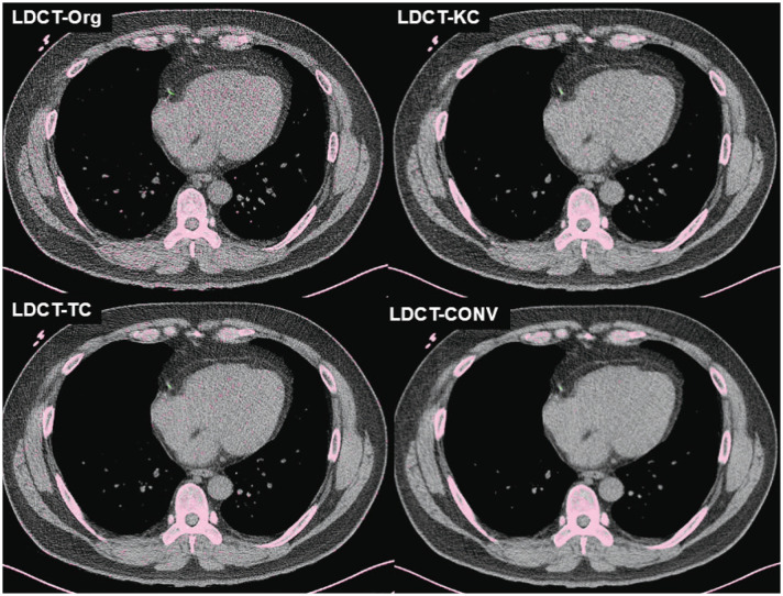
Materials and methods: A total of 225 pairs of LDCT and calcium scoring CT (CSCT) images scanned at 120 kVp and acquired from the same patient within a 6-month interval were retrospectively collected from four institutions. Image conversion was performed for LDCT images using proprietary software programs to simulate conventional CSCT. This process included 1) deep learning-based kernel conversion of low-dose, high-frequency, sharp kernels to simulate standard-dose, low-frequency kernels, and 2) thickness conversion using the raysum method to convert 1-mm or 1.25-mm thickness images to 3-mm thickness. Automated Agaston scoring was conducted on the LDCT scans before (LDCT-Orgauto) and after the image conversion (LDCT-CONVauto). Manual scoring was performed on the CSCT images (CSCTmanual) and used as a reference standard. The accuracy of automated Agaston scores and risk severity categorization based on the automated scoring on LDCT scans was analyzed compared to the reference standard, using the Bland-Altman analysis, concordance correlation coefficient (CCC), and weighted kappa (κ) statistic.
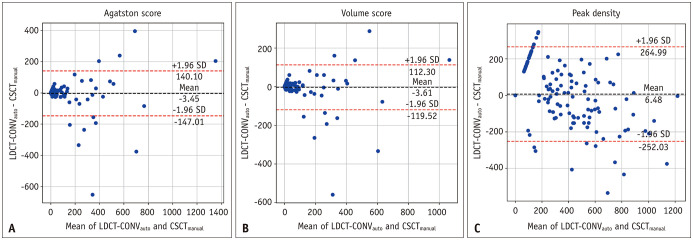
Results: LDCT-CONVauto demonstrated a reduced bias for Agaston score, compared with CSCTmanual, than LDCT-Orgauto did (-3.45 vs. 206.7). LDCT-CONVauto showed a higher CCC than LDCT-Orgauto did (0.881 [95% confidence interval {CI}, 0.750-0.960] vs. 0.269 [95% CI, 0.129-0.430]). In terms of risk category assignment, LDCT-Orgauto exhibited poor agreement with CSCTmanual (weighted κ = 0.115 [95% CI, 0.082-0.154]), whereas LDCT-CONVauto achieved good agreement (weighted κ = 0.792 [95% CI, 0.731-0.847]).
Conclusion: Deep learning-based conversion of LDCT images originally obtained with thin slices and a sharp kernel can enhance the accuracy of automated coronary artery calcium score measurement using the images.
期刊介绍:
The inaugural issue of the Korean J Radiol came out in March 2000. Our journal aims to produce and propagate knowledge on radiologic imaging and related sciences.
A unique feature of the articles published in the Journal will be their reflection of global trends in radiology combined with an East-Asian perspective. Geographic differences in disease prevalence will be reflected in the contents of papers, and this will serve to enrich our body of knowledge.
World''s outstanding radiologists from many countries are serving as editorial board of our journal.

 求助内容:
求助内容: 应助结果提醒方式:


