{"title":"Erratum to “Recent Progress in Quantitative Analysis of Self-Assembled Peptides”","authors":"","doi":"10.1002/EXP.20250016","DOIUrl":null,"url":null,"abstract":"<p>X. Cai, W. Xu, C. Ren, et al. Recent Progress in Quantitative Analysis of Self-Assembled Peptides. <i>Exploration</i> 4 (2024): 20230064.</p><p>In our manuscript titled “Recent Progress in Quantitative Analysis of Self-Assembled Peptides,” there was a mistake regarding the citation of Figure 10A–F. The true source of these data is from the publication https://doi.org/10.1021/acs.nanolett.1c03683. However, in Figure 10B, the first image in the first row is identical to the fourth image, and the first image in the third row also exhibits similarity to the fourth image. This related work was from Ye's group other than our own study. Thus, we have replaced the relevant Figure 10A–F with the data from another reference (https://doi.org/10.1021/jacs.9b03649). This revision will not affect the conclusions of this manuscript. This corrigendum corrects this error by providing the following revised section.</p><p>For multimodal imaging induced by enzyme catalysis, Ye's group designed an activable FLI/PET bimodal nanoprobe (P-CyFF-Gd) for bimodal imaging of ALP enzyme activity.<sup>[112]</sup> As shown in Figure 10A, an overexpressed enzyme, alkaline phosphatase (ALP), on the cell membrane was employed to activate the fluorescence by dephosphorylation, after which P-CyFF-Gd in situ co-assembled into nanoparticles, simultaneously switching on FLI signals and enhancing MRI imaging. In vivo bimodal imaging results showed that after intravenous injection of P-CyFF-Gd, fluorescence (Figure 10B,D) in the tumor region reached the highest level at 2 h, which was approximately 2.8 folds as high as that pretreated with ALP inhibitor (Na<sub>3</sub>VO<sub>4</sub>). Akin to FLI, MRI imaging (Figure 10C and E) displayed maximum signal enhancement (%SE) at 4 h in tumors upon injection of P-CyFF-Gd. Moreover, the distribution in tumor was higher than in other organs (Figure 10F). These studies corroborated that concomitant NIR FLI and MRI imaging of self-assembly of P-CyFF-Gd are capable of efficiently detection of enzyme activity in vivo.</p><p>We apologize for this error.</p>","PeriodicalId":72997,"journal":{"name":"Exploration (Beijing, China)","volume":"5 3","pages":""},"PeriodicalIF":22.5000,"publicationDate":"2025-02-04","publicationTypes":"Journal Article","fieldsOfStudy":null,"isOpenAccess":false,"openAccessPdf":"https://onlinelibrary.wiley.com/doi/epdf/10.1002/EXP.20250016","citationCount":"0","resultStr":null,"platform":"Semanticscholar","paperid":null,"PeriodicalName":"Exploration (Beijing, China)","FirstCategoryId":"1085","ListUrlMain":"https://onlinelibrary.wiley.com/doi/10.1002/EXP.20250016","RegionNum":0,"RegionCategory":null,"ArticlePicture":[],"TitleCN":null,"AbstractTextCN":null,"PMCID":null,"EPubDate":"","PubModel":"","JCR":"","JCRName":"","Score":null,"Total":0}
引用次数: 0
Abstract
X. Cai, W. Xu, C. Ren, et al. Recent Progress in Quantitative Analysis of Self-Assembled Peptides. Exploration 4 (2024): 20230064.
In our manuscript titled “Recent Progress in Quantitative Analysis of Self-Assembled Peptides,” there was a mistake regarding the citation of Figure 10A–F. The true source of these data is from the publication https://doi.org/10.1021/acs.nanolett.1c03683. However, in Figure 10B, the first image in the first row is identical to the fourth image, and the first image in the third row also exhibits similarity to the fourth image. This related work was from Ye's group other than our own study. Thus, we have replaced the relevant Figure 10A–F with the data from another reference (https://doi.org/10.1021/jacs.9b03649). This revision will not affect the conclusions of this manuscript. This corrigendum corrects this error by providing the following revised section.
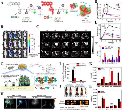
For multimodal imaging induced by enzyme catalysis, Ye's group designed an activable FLI/PET bimodal nanoprobe (P-CyFF-Gd) for bimodal imaging of ALP enzyme activity.[112] As shown in Figure 10A, an overexpressed enzyme, alkaline phosphatase (ALP), on the cell membrane was employed to activate the fluorescence by dephosphorylation, after which P-CyFF-Gd in situ co-assembled into nanoparticles, simultaneously switching on FLI signals and enhancing MRI imaging. In vivo bimodal imaging results showed that after intravenous injection of P-CyFF-Gd, fluorescence (Figure 10B,D) in the tumor region reached the highest level at 2 h, which was approximately 2.8 folds as high as that pretreated with ALP inhibitor (Na3VO4). Akin to FLI, MRI imaging (Figure 10C and E) displayed maximum signal enhancement (%SE) at 4 h in tumors upon injection of P-CyFF-Gd. Moreover, the distribution in tumor was higher than in other organs (Figure 10F). These studies corroborated that concomitant NIR FLI and MRI imaging of self-assembly of P-CyFF-Gd are capable of efficiently detection of enzyme activity in vivo.

 求助内容:
求助内容: 应助结果提醒方式:


