{"title":"Retinoblastoma in a Young Nigerian Girl: A Case Report From ECWA Eye Hospital, Kano.","authors":"Emamoke Atima-Ayeni, Ayodele Jacob Orugun, Ugbede Idakwo, Oyeronke Komolafe, Mayor Orezime Atima, Akinfenwa Taoheed Atanda, Waziri Garba Dahiru, Sani Kamarudeen Owolabi, Eisuke Shimizu, Nakayama Shintaro, Emmanuel Oluwadare Balogun, Emeka John Dingwoke","doi":"10.1155/crop/1733830","DOIUrl":null,"url":null,"abstract":"<p><p><b>Objective:</b> This report details the case of a 9-year-old Nigerian girl presenting with proptosis and a fungating ocular mass, which was histologically confirmed as retinoblastoma following exenteration. <b>Introduction:</b> Retinoblastoma is the most common pediatric intraocular malignancy, predominantly affecting infants and children under the age of 5, with leukocoria being the most frequent presenting symptom. The occurrence of retinoblastoma in older children is rare and often associated with atypical presentations. <b>Case Summary:</b> A 9-year-old Nigerian girl presented with a 1-year history of progressive left eye symptoms, including redness, pain, decreased vision, and proptosis. Examination revealed a large, fungating ocular mass with no light perception. Imaging studies (ultrasound B-scan and CT scan) confirmed extensive vitreous infiltration and optic nerve involvement. Histopathological analysis postmodified exenteration confirmed retinoblastoma. The patient clinically tolerated a modified exenteration and the first two of six planned cycles of systemic chemotherapy (vincristine, etoposide, and carboplatin). Unfortunately, she died during the second month of follow-up. The recurrence was characterized by progressive worsening of systemic symptoms and preauricular lymphadenopathy, likely indicating metastatic spread. <b>Conclusion:</b> This case highlights the aggressive progression of advanced retinoblastoma and the consequences of delayed presentation in resource-limited settings. Although the patient demonstrated an initial positive response, clinically tolerating modified exenteration and two cycles of systemic chemotherapy, she rapidly succumbed to the disease. This underscores the critical need for early diagnosis, prompt referral, and improved access to specialized care to enhance outcomes in similar contexts.</p>","PeriodicalId":9603,"journal":{"name":"Case Reports in Ophthalmological Medicine","volume":"2025 ","pages":"1733830"},"PeriodicalIF":0.4000,"publicationDate":"2025-06-08","publicationTypes":"Journal Article","fieldsOfStudy":null,"isOpenAccess":false,"openAccessPdf":"https://www.ncbi.nlm.nih.gov/pmc/articles/PMC12170086/pdf/","citationCount":"0","resultStr":null,"platform":"Semanticscholar","paperid":null,"PeriodicalName":"Case Reports in Ophthalmological Medicine","FirstCategoryId":"1085","ListUrlMain":"https://doi.org/10.1155/crop/1733830","RegionNum":0,"RegionCategory":null,"ArticlePicture":[],"TitleCN":null,"AbstractTextCN":null,"PMCID":null,"EPubDate":"2025/1/1 0:00:00","PubModel":"eCollection","JCR":"Q4","JCRName":"OPHTHALMOLOGY","Score":null,"Total":0}
引用次数: 0
Abstract
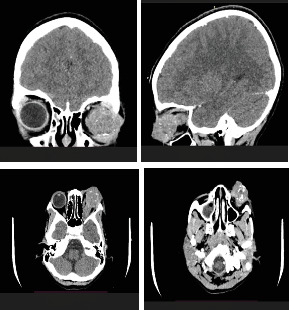
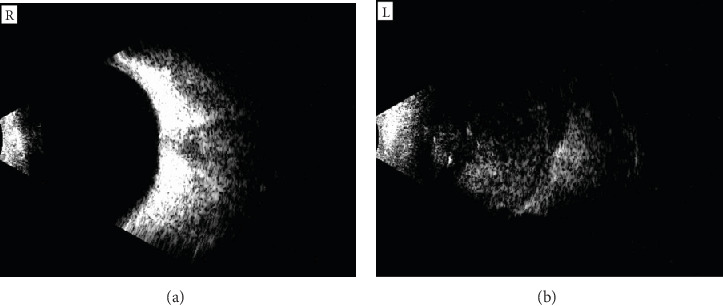
Objective: This report details the case of a 9-year-old Nigerian girl presenting with proptosis and a fungating ocular mass, which was histologically confirmed as retinoblastoma following exenteration. Introduction: Retinoblastoma is the most common pediatric intraocular malignancy, predominantly affecting infants and children under the age of 5, with leukocoria being the most frequent presenting symptom. The occurrence of retinoblastoma in older children is rare and often associated with atypical presentations. Case Summary: A 9-year-old Nigerian girl presented with a 1-year history of progressive left eye symptoms, including redness, pain, decreased vision, and proptosis. Examination revealed a large, fungating ocular mass with no light perception. Imaging studies (ultrasound B-scan and CT scan) confirmed extensive vitreous infiltration and optic nerve involvement. Histopathological analysis postmodified exenteration confirmed retinoblastoma. The patient clinically tolerated a modified exenteration and the first two of six planned cycles of systemic chemotherapy (vincristine, etoposide, and carboplatin). Unfortunately, she died during the second month of follow-up. The recurrence was characterized by progressive worsening of systemic symptoms and preauricular lymphadenopathy, likely indicating metastatic spread. Conclusion: This case highlights the aggressive progression of advanced retinoblastoma and the consequences of delayed presentation in resource-limited settings. Although the patient demonstrated an initial positive response, clinically tolerating modified exenteration and two cycles of systemic chemotherapy, she rapidly succumbed to the disease. This underscores the critical need for early diagnosis, prompt referral, and improved access to specialized care to enhance outcomes in similar contexts.


 求助内容:
求助内容: 应助结果提醒方式:


