Christian Nelles, Philip Rauen, Franziska Meyer, Anja Dobrostal, Pia Lena Niederau, Hasan Zaytoun, Mathilda Weisthoff, Pascale Bernard, Carola Heneweer, Thomas Dratsch, David Maintz, Jonathan Kottlors, Nicole Kreuzberg, Nils Große Hokamp, David Zopfs, Thorsten Persigehl, Simon Lennartz
{"title":"Spectral CT imaging for assessment of metastases in melanoma patients: multi-reader evaluation.","authors":"Christian Nelles, Philip Rauen, Franziska Meyer, Anja Dobrostal, Pia Lena Niederau, Hasan Zaytoun, Mathilda Weisthoff, Pascale Bernard, Carola Heneweer, Thomas Dratsch, David Maintz, Jonathan Kottlors, Nicole Kreuzberg, Nils Große Hokamp, David Zopfs, Thorsten Persigehl, Simon Lennartz","doi":"10.1186/s40644-025-00889-7","DOIUrl":null,"url":null,"abstract":"<p><strong>Background: </strong>Pilot studies have indicated diagnostic benefits from using dual-energy CT (DECT) for staging and follow-up of melanoma patients. The purpose of this study was to investigate the sensitivity, specificity and qualitative assessment of spectral image reconstructions for metastases in melanoma patients in a large-scale, multi-reader evaluation.</p><p><strong>Methods: </strong>In total, 308 patients with melanoma, 95 patients with metastases and a control group of 213 patients without metastases, who underwent oncologic staging CT of the chest, abdomen and pelvis on a dual-layer dual-energy CT system (dlDECT) were retrospectively included. Conventional images (CI), color-coded iodine overlays (IO) and virtual monoenergetic images at 40 keV (VMI<sub>40keV</sub>) were reconstructed. 6 radiologists (3 experienced with 6 to 9 years and 3 less experienced with 2 to 4 years of experience) read all cases in a CI-based session, and a session based on a combination of CI, IO and VMI<sub>40keV</sub>. Readers were asked to determine presence of metastases in specific tissues in a binary fashion and to indicate diagnostic certainty and lesion delineation on 5-point Likert scales.</p><p><strong>Results: </strong>Sensitivity for detection of metastases in the skeletal muscle and peritoneum was significantly higher for the spectral assessment (for skeletal muscle 70% vs. 61%; for peritoneum 76% vs. 62%, both: p < 0.05). For subcutaneous metastases, there was a significant increase in specificity (92% vs. 89%, p < 0.05), however accompanied with a significant decrease in sensitivity (79% vs. 85%, p < 0.05). Diagnostic certainty was rated significantly higher for spectral images than CI in all (6/6) of the assessed tissues, whereas improvements in lesion delineation were noted for the skeletal muscle, the subcutaneous tissue and the pancreas.</p><p><strong>Conclusions: </strong>We found that in melanoma patients, the benefit of dlDECT-derived spectral reconstructions depends on the assessed tissue. While assessment of skeletal muscle and peritoneal metastases was significantly improved, low or absent iodine uptake of subcutaneous lesions led to false negatives and a consecutive decrease in sensitivity.</p>","PeriodicalId":9548,"journal":{"name":"Cancer Imaging","volume":"25 1","pages":"74"},"PeriodicalIF":3.5000,"publicationDate":"2025-06-16","publicationTypes":"Journal Article","fieldsOfStudy":null,"isOpenAccess":false,"openAccessPdf":"https://www.ncbi.nlm.nih.gov/pmc/articles/PMC12168337/pdf/","citationCount":"0","resultStr":null,"platform":"Semanticscholar","paperid":null,"PeriodicalName":"Cancer Imaging","FirstCategoryId":"3","ListUrlMain":"https://doi.org/10.1186/s40644-025-00889-7","RegionNum":2,"RegionCategory":"医学","ArticlePicture":[],"TitleCN":null,"AbstractTextCN":null,"PMCID":null,"EPubDate":"","PubModel":"","JCR":"Q2","JCRName":"ONCOLOGY","Score":null,"Total":0}
引用次数: 0
Abstract
Background: Pilot studies have indicated diagnostic benefits from using dual-energy CT (DECT) for staging and follow-up of melanoma patients. The purpose of this study was to investigate the sensitivity, specificity and qualitative assessment of spectral image reconstructions for metastases in melanoma patients in a large-scale, multi-reader evaluation.
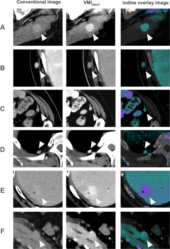
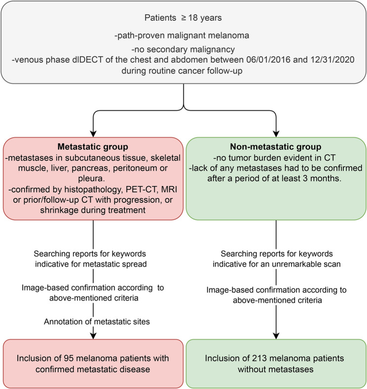
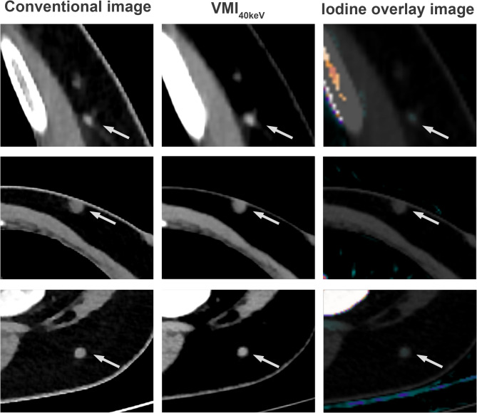
Methods: In total, 308 patients with melanoma, 95 patients with metastases and a control group of 213 patients without metastases, who underwent oncologic staging CT of the chest, abdomen and pelvis on a dual-layer dual-energy CT system (dlDECT) were retrospectively included. Conventional images (CI), color-coded iodine overlays (IO) and virtual monoenergetic images at 40 keV (VMI40keV) were reconstructed. 6 radiologists (3 experienced with 6 to 9 years and 3 less experienced with 2 to 4 years of experience) read all cases in a CI-based session, and a session based on a combination of CI, IO and VMI40keV. Readers were asked to determine presence of metastases in specific tissues in a binary fashion and to indicate diagnostic certainty and lesion delineation on 5-point Likert scales.
Results: Sensitivity for detection of metastases in the skeletal muscle and peritoneum was significantly higher for the spectral assessment (for skeletal muscle 70% vs. 61%; for peritoneum 76% vs. 62%, both: p < 0.05). For subcutaneous metastases, there was a significant increase in specificity (92% vs. 89%, p < 0.05), however accompanied with a significant decrease in sensitivity (79% vs. 85%, p < 0.05). Diagnostic certainty was rated significantly higher for spectral images than CI in all (6/6) of the assessed tissues, whereas improvements in lesion delineation were noted for the skeletal muscle, the subcutaneous tissue and the pancreas.
Conclusions: We found that in melanoma patients, the benefit of dlDECT-derived spectral reconstructions depends on the assessed tissue. While assessment of skeletal muscle and peritoneal metastases was significantly improved, low or absent iodine uptake of subcutaneous lesions led to false negatives and a consecutive decrease in sensitivity.
背景:前期研究表明,双能CT (DECT)对黑色素瘤患者的分期和随访有诊断价值。本研究的目的是在大规模、多阅读器评估中探讨光谱图像重建对黑色素瘤患者转移的敏感性、特异性和定性评估。方法:回顾性分析308例黑色素瘤患者、95例转移患者和213例无转移患者,均行双层双能CT系统(dlDECT)胸腹骨盆肿瘤分期CT检查。重建常规图像(CI)、彩色编码碘叠加图像(IO)和虚拟单能图像(VMI40keV)。6名放射科医生(3名有6至9年经验,3名经验较少,有2至4年经验)在基于CI的会议上阅读所有病例,以及基于CI, IO和VMI40keV组合的会议。读者被要求以二元方式确定特定组织中转移的存在,并在5点李克特量表上指示诊断的确定性和病变的描述。结果:在光谱评估中,骨骼肌和腹膜转移检测的敏感性明显更高(骨骼肌70% vs. 61%;结论:我们发现在黑色素瘤患者中,dldect衍生的光谱重建的益处取决于评估的组织。虽然骨骼肌和腹膜转移的评估得到了显著改善,但皮下病变的碘摄取低或无碘摄取导致假阴性和敏感性持续下降。
Cancer ImagingONCOLOGY-RADIOLOGY, NUCLEAR MEDICINE & MEDICAL IMAGING
CiteScore
7.00
自引率
0.00%
发文量
66
审稿时长
>12 weeks
期刊介绍:
Cancer Imaging is an open access, peer-reviewed journal publishing original articles, reviews and editorials written by expert international radiologists working in oncology.
The journal encompasses CT, MR, PET, ultrasound, radionuclide and multimodal imaging in all kinds of malignant tumours, plus new developments, techniques and innovations. Topics of interest include:
Breast Imaging
Chest
Complications of treatment
Ear, Nose & Throat
Gastrointestinal
Hepatobiliary & Pancreatic
Imaging biomarkers
Interventional
Lymphoma
Measurement of tumour response
Molecular functional imaging
Musculoskeletal
Neuro oncology
Nuclear Medicine
Paediatric.

 求助内容:
求助内容: 应助结果提醒方式:


